

Moderate cardiac-selective overexpression of angiotensin II type 2 receptor protects cardiac functions from ischaemic injury
- PMID: 21967903
- PMCID: PMC3619662
- DOI: 10.1113/expphysiol.2011.060673
Moderate cardiac-selective overexpression of angiotensin II type 2 receptor protects cardiac functions from ischaemic injury
Abstract
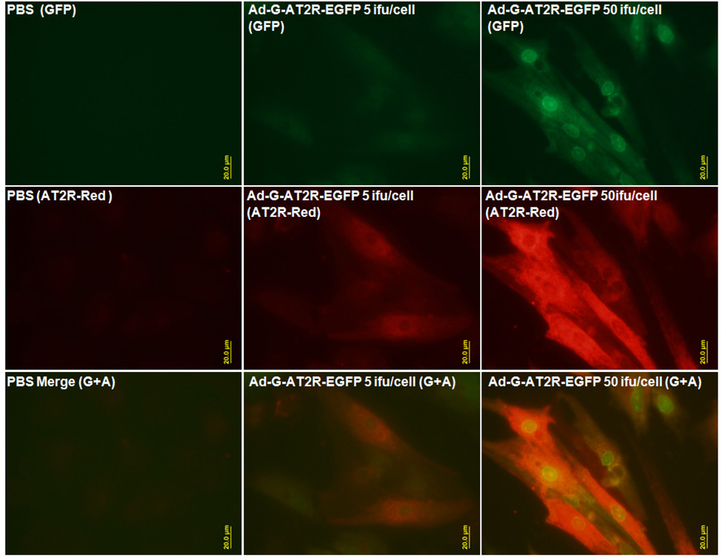
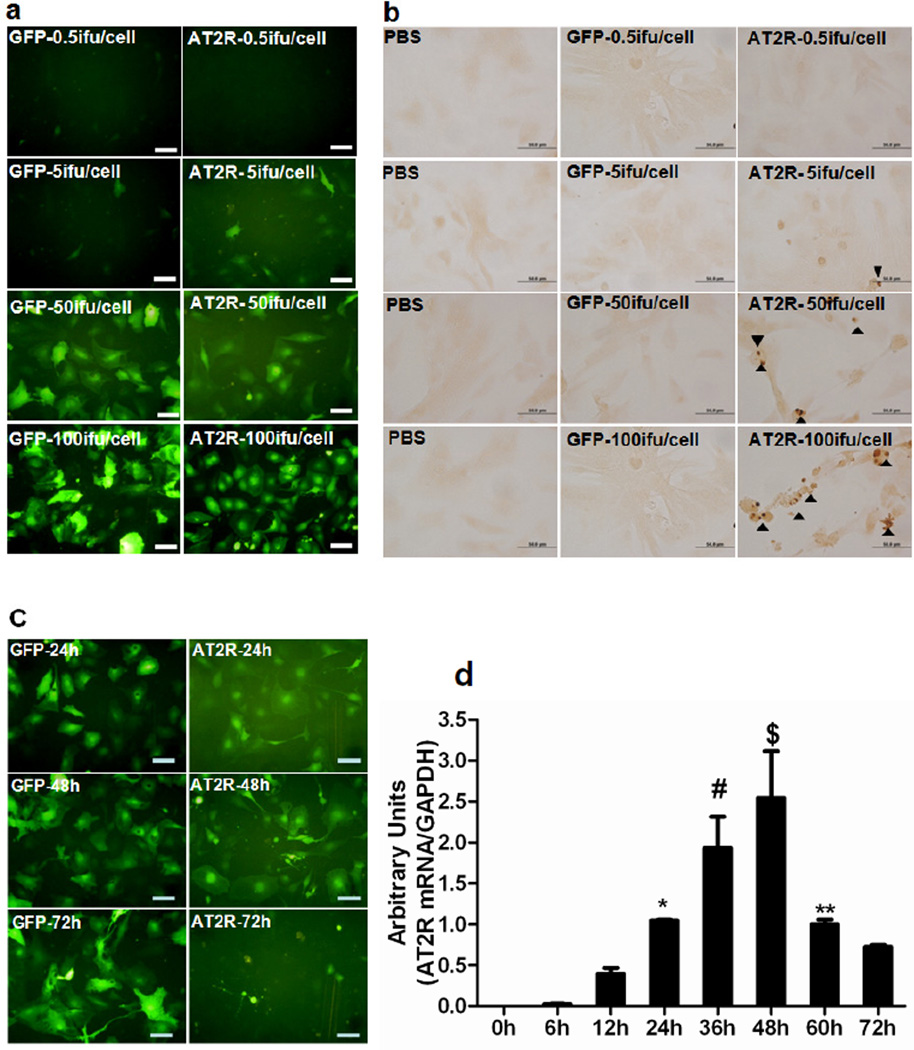
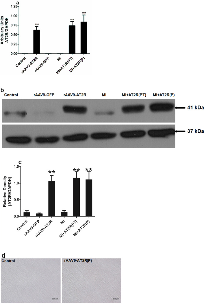
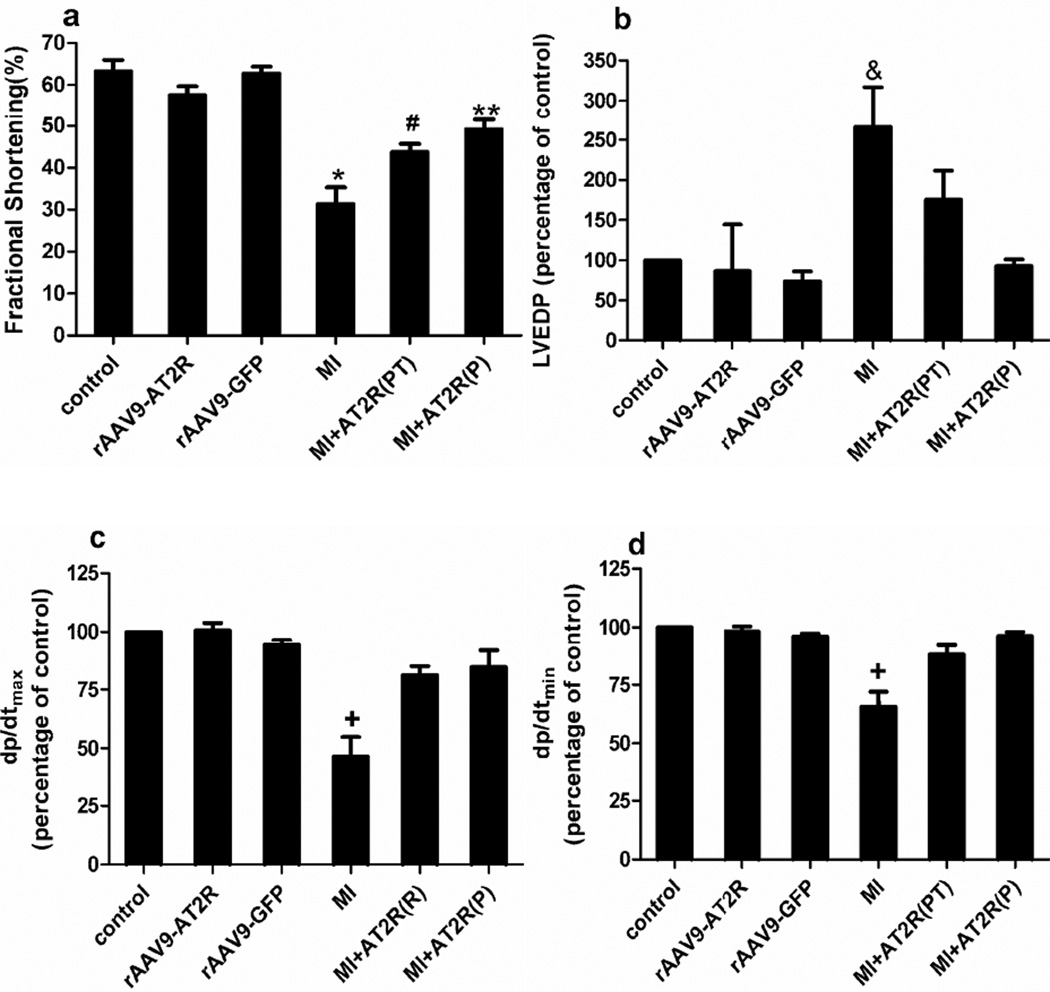
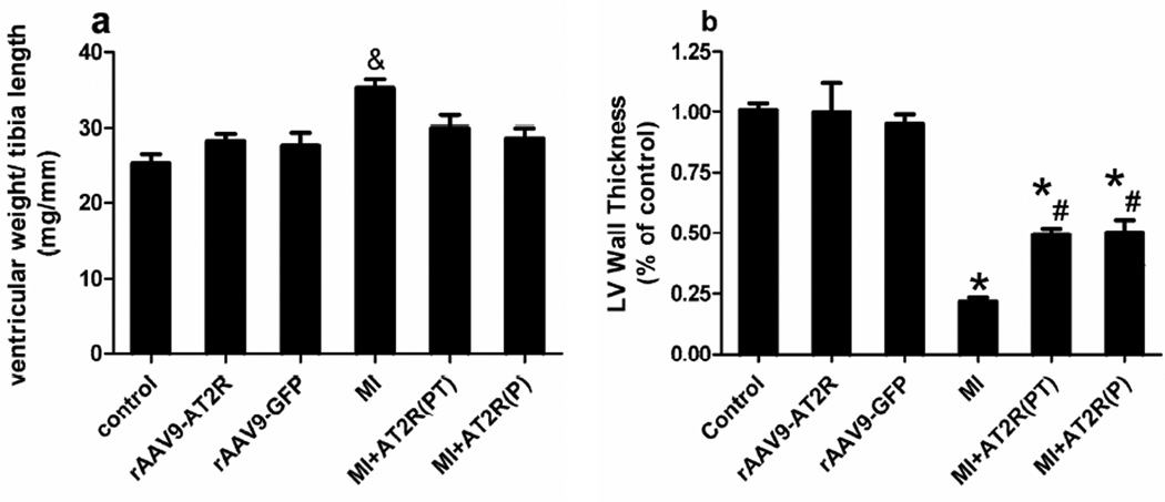
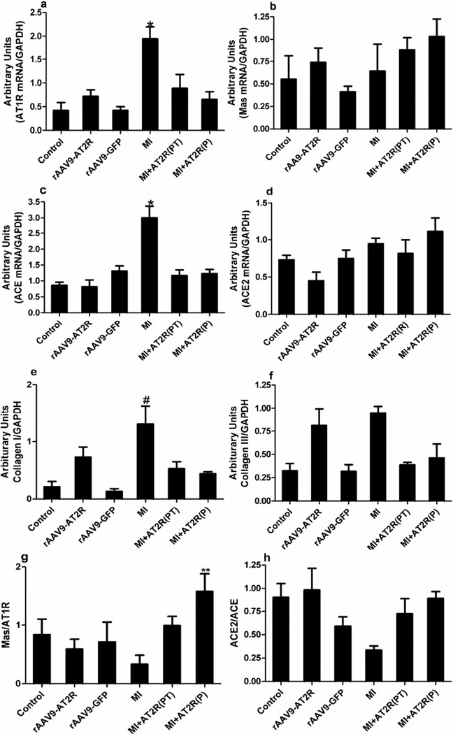
We hypothesized that moderate cardiac-selective overexpression of the angiotensin II type 2 receptor (AT2R) would protect the myocardium from ischaemic injury after a myocardial infarction (MI) induced by coronary artery ligation. For in vitro studies, adenoviral vector expressing genomic DNA of AT2R and enhanced green fluorescence protein (EGFP) was used to overexpress AT2R in rat neonatal cardiac myocytes. Expression of AT2R, measured by real-time PCR and immunostaining, demonstrated efficient transduction of AT2R in a dose-dependent pattern. The AT2R constitutively induced apoptosis in rat neonatal cardiac myocytes in dose-dependent patterns. For in vivo studies, 4 × 10(10) vector genomes (vg) of recombinant adeno-associated virus serotype 9 (rAAV9)-chicken β actin promoter-AT2R was injected into the left ventricle of 5-day-old Sprague-Dawley rats. At 6 weeks of age, hearts were harvested and expression of AT2R determined by real-time PCR and Western blotting. Expression was increased onefold over control hearts, and no apoptosis was detected. Two subsequent in vivo studies were performed. In a prevention study, 4 × 10(10) vg of rAAV9-CBA-AT2R was injected into the left ventricle of 5-day-old Sprague-Dawley rats and MI was induced at 6 weeks of age. For a post-treatment study, 4 × 10(10) vg of rAAV9-CBA-AT2R was administrated to the peri-infarcted myocardium area immediately after MI in 6-week-old animals. For both in vivo studies, cardiac functions were assessed using echocardiography and haemodynamic measurements 4 weeks after coronary artery ligation. In the in vivo studies, the rats subjected to MI showed significant decreases in fractional shortening and rate of change of left ventricular pressure, with increased left ventricular end-diastolic pressure and ventricular hypertrophy. For the prevention study, the moderate cardiac-selective overexpression of AT2R attenuated these MI-induced impairments and also caused a decrease in ventricular wall thinning. In the post-treatment study, the overexpression of AT2R partly reversed the MI-induced cardiac dysfunction. Myocardial infarction also induced the upregulation of angiotensin II type 1 receptor, angiotensin-converting enzyme and collagen I mRNA expression, all of which were attenuated by the overexpression of AT2R. It is concluded that moderate cardiac-selective overexpression of AT2R protects heart function from ischaemic injury, which may be mediated, at least in part, through modulation of components of the cardiac renin-angiotensin system and collagen levels in the myocardium.
Figures
Comment in
-
Is angiotensin II type 2 receptor a new therapeutic target for cardiovascular disease?Exp Physiol. 2014 Jul;99(7):933-4. doi: 10.1113/expphysiol.2014.080705. Exp Physiol. 2014. PMID: 24987019 No abstract available.
Similar articles
-
Effects of cardiac overexpression of the angiotensin II type 2 receptor on remodeling and dysfunction in mice post-myocardial infarction.Hypertension. 2014 Jun;63(6):1251-9. doi: 10.1161/HYPERTENSIONAHA.114.03247. Epub 2014 Apr 14. Hypertension. 2014. PMID: 24732892 Free PMC article.
-
Lentivirus-mediated overexpression of angiotensin-(1-7) attenuated ischaemia-induced cardiac pathophysiology.Exp Physiol. 2011 Sep;96(9):863-74. doi: 10.1113/expphysiol.2011.056994. Epub 2011 Jun 17. Exp Physiol. 2011. PMID: 21685447 Free PMC article.
-
Prevention of cardiac hypertrophy by angiotensin II type-2 receptor gene transfer.Hypertension. 2004 Jun;43(6):1233-8. doi: 10.1161/01.HYP.0000127563.14064.FD. Hypertension. 2004. PMID: 15166182
-
Effects of angiotensin-converting enzyme inhibition and angiotensin II AT1 receptor antagonism on cardiac parameters in left ventricular hypertrophy.Am J Cardiol. 1997 Aug 4;80(3A):110A-117A. doi: 10.1016/s0002-9149(97)00465-7. Am J Cardiol. 1997. PMID: 9293963 Review.
-
AT2 receptors targeting cardiac protection post-myocardial infarction.Curr Hypertens Rep. 2014 Jul;16(7):441. doi: 10.1007/s11906-014-0441-0. Curr Hypertens Rep. 2014. PMID: 24792092 Review.
Cited by
-
The Protective Effects of Compound 21 and Valsartan in Isoproterenol-Induced Myocardial Injury in Rats.Cardiovasc Toxicol. 2021 Jan;21(1):17-28. doi: 10.1007/s12012-020-09590-6. Epub 2020 Jul 9. Cardiovasc Toxicol. 2021. PMID: 32648158
-
Effects of cardiac overexpression of the angiotensin II type 2 receptor on remodeling and dysfunction in mice post-myocardial infarction.Hypertension. 2014 Jun;63(6):1251-9. doi: 10.1161/HYPERTENSIONAHA.114.03247. Epub 2014 Apr 14. Hypertension. 2014. PMID: 24732892 Free PMC article.
-
The Impact of the Renin-Angiotensin-Aldosterone System on Inflammation, Coagulation, and Atherothrombotic Complications, and to Aggravated COVID-19.Front Pharmacol. 2021 Jun 17;12:640185. doi: 10.3389/fphar.2021.640185. eCollection 2021. Front Pharmacol. 2021. PMID: 34220496 Free PMC article. Review.
-
Withaferin A attenuates ovarian cancer-induced cardiac cachexia.PLoS One. 2020 Jul 28;15(7):e0236680. doi: 10.1371/journal.pone.0236680. eCollection 2020. PLoS One. 2020. PMID: 32722688 Free PMC article.
-
Breakthrough: a first-in-class virtual simulator for dose optimization of ACE inhibitors in translational cardiovascular medicine.Sci Rep. 2023 Feb 26;13(1):3300. doi: 10.1038/s41598-023-30453-x. Sci Rep. 2023. PMID: 36843132 Free PMC article.
References
-
- Anavekar NS, Solomon SD. Angiotensin II receptor blockade and ventricular remodelling. J.Renin Angiotensin Aldosterone Syst. 2005;6:43–48. - PubMed
-
- Booz GW, Baker KM. Role of type 1 and type 2 angiotensin receptors in angiotensin II-induced cardiomyocyte hypertrophy. Hypertension. 1996;28:635–640. - PubMed
-
- Booz GW, Dostal DE, Baker KM. Paracrine actions of cardiac fibroblasts on cardiomyocytes: implications for the cardiac renin-angiotensin system. Am.J.Cardiol. 1999;83:44H–47H. - PubMed
-
- Castro CH, Santos RA, Ferreira AJ, Bader M, Alenina N, Almeida AP. Evidence for a functional interaction of the angiotensin-(1–7) receptor Mas with AT1 and AT2 receptors in the mouse heart. Hypertension. 2005;46:937–942. - PubMed
-
- D'Amore A, Black MJ, Thomas WG. The angiotensin II type 2 receptor causes constitutive growth of cardiomyocytes and does not antagonize angiotensin II type 1 receptormediated hypertrophy. Hypertension. 2005;46:1347–1354. - PubMed
MeSH terms
Substances
Grants and funding
LinkOut - more resources
Full Text Sources
Other Literature Sources