. 2011 Dec 10;10(12):1243-51.
doi: 10.1016/j.dnarep.2011.09.013. Epub 2011 Oct 2.

A role for SUMO in nucleotide excision repair

Affiliations

A role for SUMO in nucleotide excision repair

Hannah R Silver et al. DNA Repair (Amst). .

Abstract

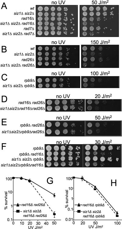
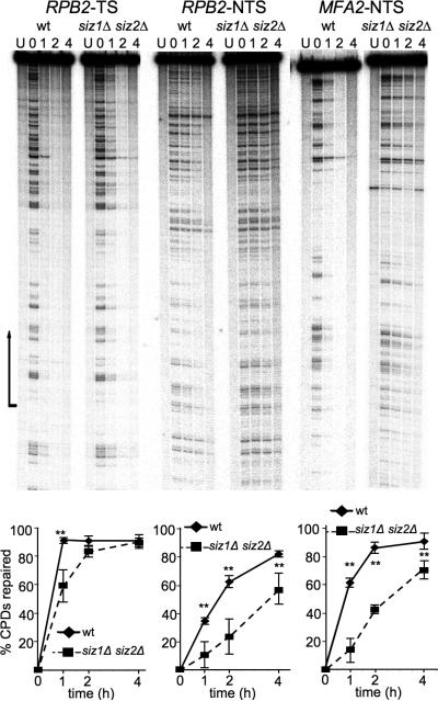
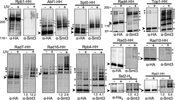
The two Siz/PIAS SUMO E3 ligases Siz1 and Siz2 are responsible for the vast majority of sumoylation in Saccharomyces cerevisiae. We found that siz1Δ siz2Δ mutants are sensitive to ultra-violet (UV) light. Epistasis analysis showed that the SIZ genes act in the nucleotide excision repair (NER) pathway, and suggested that they participate both in global genome repair (GGR) and in the Rpb9-dependent subpathway of transcription-coupled repair (TCR), but have minimal role in Rad26-dependent TCR. Quantitative analysis of NER at the single-nucleotide level showed that siz1Δ siz2Δ is deficient in repair of both the transcribed and non-transcribed strands of the DNA. These experiments confirmed that the SIZ genes participate in GGR. Their role in TCR remains unclear. It has been reported previously that mutants deficient for the SUMO conjugating enzyme Ubc9 contain reduced levels of Rad4, the yeast homolog of human XPC. However, our experiments do not support the conclusion that SUMO conjugation affects Rad4 levels. We found that several factors that participate in NER are sumoylated, including Rad4, Rad16, Rad7, Rad1, Rad10, Ssl2, Rad3, and Rpb4. Although Rad16 was heavily sumoylated, elimination of the major SUMO attachment sites in Rad16 had no detectable effect on UV resistance or removal of DNA lesions. SUMO attachment to most of these NER factors was significantly increased by DNA damage. Furthermore, SUMO-modified Rad4 accumulated in NER mutants that block the pathway downstream of Rad4, suggesting that SUMO becomes attached to Rad4 at a specific point during its functional cycle. Collectively, these results suggest that SIZ-dependent sumoylation may modulate the activity of multiple proteins to promote efficient NER.


Figures

Fig. 1
Fig. 1
Epistasis between core NER genes and siz1Δ siz2Δ. (A, B) The indicated mutants were plated, UV irradiated at indicated intensities, and allowed to grow for three days in the dark before counting surviving colonies. Values represent averages of three independent experiments and error bars represent standard deviations. (C, D) Overnight cultures of indicated genotypes were serially diluted (10-fold), spotted onto YPD, UV irradiated at indicated doses and incubated for 2-3 days in the dark at 30°C.
Fig. 2
Fig. 2
Siz1 and Siz2 play a role in Rad16- and Rpb9-mediated NER. (A-F) Spotting of indicated mutants was completed as in Fig. 1. (G, H) Survival analyses of indicated mutants were performed as in Fig. 1.
Fig. 3
Fig. 3
siz1Δ siz2Δ shows delayed repair of CPDs. (Top) Representative autoradiograms showing repair of the indicated strands of the indicated genes in wt and siz1Δ siz2Δ. Lanes are DNA samples from untreated cells (NT) and UV-irradiated cells following different times of repair incubation (hours) as indicated over each lane. Arrow indicates transcription start site and direction of transcription. For MFA2-NTS, the wt and siz1Δ siz2Δ samples were taken from different gels. (Bottom) Plots of CPD repair for the genes and strands shown above. For TS, the region of quantification included the entire transcribed region; for NTS, the whole gel was quantified. Error bars represent standard deviations of three independent experiments. Two asterisks represents p<0.01.
Fig. 4
Fig. 4
NER factors modified by SUMO. His8-, or His8- and HA-tagged versions of indicated proteins from mock- and UV-treated wt strains were purified by Ni-NTA affinity chromatography and analyzed by SDS-PAGE and immunoblotting with Abs against Smt3 (right of each set), and against the HA epitope or His5, as indicated (left of each set). Cells were incubated in YPD for 30 m post-UV treatment. Ab binding in the top five sets of blots was detected using chemiluminescence, while double-label fluorescent detection was used in bottom seven blots. Arrowheads indicate unsumoylated tagged proteins. (There are two for Spt5, which is partially hyperphosphorylated.) Open circles designate sumoylated species. In some samples, sumoylated species are also visible on the HA blot. Asterisks indicate cross-reacting bands. Numbers below the lanes represent relative levels of protein sumoylation, where mock-treated was set at 1. HH, –His8-HA. Lines between HA and Smt3 blots indicate how these blots align. For the top row of blots, the HA and Smt3 blots were performed on different filters (using the same samples), so alignments are approximate. Markers are Bio-Rad broad range markers. Three sets of lanes (Rpb1, Abf1, and Spt5), (Rad4 and Top1), and (Rad7, Rad16, and Rpb4) show the same protein size range from the same exposure(s) of the same blot(s), and so the background and markers for these lanes can be compared to each other.
Fig. 5
Fig. 5
Analysis of Rad16 sumoylation. (A) Sumoylation of Rad16-HH and Rad16-HH mutants in wt cells was analyzed as in Fig. 4, using chemiluminescent detection. Designations are as in Fig. 4. The same 4 sumoylated species indicated in Fig. 4 are indicated. Arrows on Smt3 blots indicate position of unmodified Rad16-HH. HH, His8-HA; Rad16ΔN-HH, N-terminal deletion mutant lacking the first 103 residues. Markers are Bio-Rad broad range markers. (B) Sumoylation of Rad16-HH in cells of indicated genotypes, and of SUMO attachment site mutant in wt cells, was analyzed as in Fig. 4, using fluorescent detection. Designations are as in Fig. 4. Arrows on Smt3 blots indicate position of unmodified Rad16-HH. Numbers under the lanes indicate the ratio of Smt3 signal relative to unmodified species, where all ratios are normalized to the wt UV-treated sample. Rad16K3R-HH: Rad16K62,93,103R-HH. Markers are Licor single color markers. (C) Spotting of indicated strains completed as in Fig. 1.
Fig. 6
Fig. 6
Rad4 steady state levels are not altered in SUMO pathway mutants. (A) Whole cell lysates from cells of indicated genotype were analyzed by SDS-PAGE and double-label fluorescent immunoblotting with Abs against Rad4, and PSTAIR as a loading control. Ratio of Rad4 to PSTAIR is shown, normalized to wt. Error bars represent standard deviations of 3-4 independent experiments. Two asterisks represent p<0.01. (B) Cultures of indicated genotype were grown to log phase at 22°C and shifted to 36°C for the indicated time (h), except extreme right two lanes, which were grown long-term at 30°C and grown to log phase. Whole cell lysates were analyzed as in (A) using Abs against Rad4 (top panel) and PSTAIR (not shown) or by chemiluminescent immunoblotting with an Ab against Smt3 (bottom panel). Numbers beneath the lanes indicate ratios of Rad4 to PSTAIR, with ratios normalized to the wt 0 time point. Arrowhead indicates Rad4. (C) Cultures of the indicated genotypes were treated with 50 μg/ml CHX immediately before UV or mock treatment. Cells were returned to culture containing CHX, for the indicated time (h). Whole cell lysates were analyzed as in (B). Designations as in (B).
Fig. 7
Fig. 7
Analysis of Rad4 sumoylation. Sumoylation of Rad4-HH in strains of the indicated genotypes was analyzed as in Fig. 4, using fluorescent detection. Designations as in Fig. 4. The same 2 sumoylated species indicated in Fig. 4 are indicated. Numbers under the lanes indicate normalized ratio of Smt3 signal to HA signal. In the top panel, ratios were normalized to UV-irradiated wt, while in other panels, ratios were normalized to sample with the highest ratio. rad16Δ and rad1Δ samples were from the same blot, but lanes in between were removed. For the UV-irradiated rad1Δ sample, an overexposed version of the α-HA signal is shown next to the same lane with the α-Smt3 signal overlaid.

Similar articles

Cited by

References

    1. Gareau JR, Lima CD. The SUMO pathway: emerging mechanisms that shape specificity, conjugation and recognition. Nat Rev Mol Cell Biol. 2010;11:861–871. - PMC - PubMed
    1. Geiss-Friedlander R, Melchior F. Concepts in sumoylation: a decade on. Nat Rev Mol Cell Biol. 2007;8:947–956. - PubMed
    1. Thomson TM, Guerra-Rebollo M. Ubiquitin and SUMO signalling in DNA repair. Biochem Soc Trans. 2010;38:116–131. - PubMed
    1. Wilkinson KA, Henley JM. Mechanisms, regulation and consequences of protein SUMOylation. Biochem J. 2010;428:133–145. - PMC - PubMed
    1. Ulrich HD. The SUMO system: an overview. Methods Mol Biol. 2009;497:3–16. - PubMed

Publication types

MeSH terms