. 2011 Nov;122(5):615-26.
doi: 10.1007/s00401-011-0881-4. Epub 2011 Oct 4.

Bipolar disorder type 1 and schizophrenia are accompanied by decreased density of parvalbumin- and somatostatin-positive interneurons in the parahippocampal region

Affiliations

Bipolar disorder type 1 and schizophrenia are accompanied by decreased density of parvalbumin- and somatostatin-positive interneurons in the parahippocampal region

Alice Y Wang et al. Acta Neuropathol. 2011 Nov.

Abstract

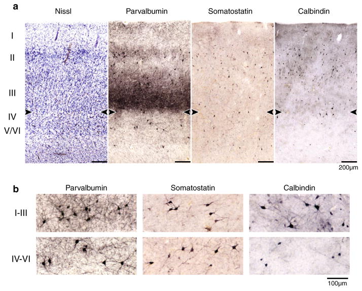
GABAergic interneurons synchronize network activities and monitor information flow. Post-mortem studies have reported decreased densities of cortical interneurons in schizophrenia (SZ) and bipolar disorder (BPD). The entorhinal cortex (EC) and the adjacent subicular regions are a hub for integration of hippocampal and cortical information, a process that is disrupted in SZ. Here we contrast and compare the density of interneuron populations in the caudal EC and subicular regions in BPD type I (BPD-I), SZ, and normal control (NC) subjects. Post-mortem human parahippocampal specimens of 13 BPD-I, 11 SZ and 17 NC subjects were used to examine the numerical density of parvalbumin-, somatostatin- or calbindin-positive interneurons. We observed a reduction in the numerical density of parvalbumin- and somatostatin-positive interneurons in the caudal EC and parasubiculum in BPD-I and SZ, but no change in the subiculum. Calbindin-positive interneuron densities were normal in all brain areas examined. The profile of decreased density was strikingly similar in BPD-I and SZ. Our results demonstrate a specific reduction of parvalbumin- and somatostatin-positive interneurons in the parahippocampal region in BPD-I and SZ, likely disrupting synchronization and integration of cortico-hippocampal circuits.


Figures

Fig. 1
Fig. 1
Overview of subiculum, presubiculum, parasubiculum and EC in a 42-year-old female NC subject. Regional borders are marked by arrowheads, boxes demarcate areas shown in subsequent figures. Neurons were counted within the entire region between two arrowheads. a Nissl stain; b PV immunocytochemistry, c SOM immunocytochemistry, d CB immunocytochemistry. Note the characteristic pattern in the PV stain. In the Nissl stain, a widening of the pyramidal cell layer between CA1 and subiculum defined the medial subicular border, whereas the narrowing of the pyramidal layer between subiculum and presubiculum defined the lateral subicular border
Fig. 2
Fig. 2
a Comparison of Nissl stain, PV immunocytochemistry, SOM immunocytochemistry and CB immunocytochemistry in the subiculum. The pyramidal cell layer (II) is located between two distinct layers. Stars denote the same blood vessel in all tissue slices. b Representative neurons. Note the different populations of CB-positive neurons
Fig. 3
Fig. 3
a Comparison of Nissl stain, PV immunocytochemistry, SOM immunocytochemistry and CB immunocytochemistry in the presubiculum, a transition zone between 3-layered hippocampus and 6-layered neocortex. The presubiculum was divided into lamina principalis externa and interna. Stars denote the same blood vessels in all tissue slices. b A lower magnification of an adjacent slice of the same subject shows the overlap between cell-dense areas in the Nissl stain and darkly stained neuropil in the PV immunocytochemistry. Arrowheads denote boundaries of the presubiculum. c Representative neurons from each lamina
Fig. 4
Fig. 4
a Comparison of Nissl stain, PV immunocytochemistry, SOM immunocytochemistry and CB immunocytochemistry in the parasubiculum. To improve contrast of layers II–VI, layer I has been overexposed in the Nissl stain. Note: the top torn part of the SOM sample was slightly rotated for better fit in the allocated space (compare to unaltered Fig. 1). b Representative neurons from layers II/III and IV–VI
Fig. 5
Fig. 5
a Comparison of Nissl stain, PV immunocytochemistry, SOM immunocytochemistry and CB immunocytochemistry in the EC. The border between layers III and IV was defined by the contrast in neuropil staining in the PV samples and the neuron-free layer IV in the Nissl stain, and is marked by arrowheads. b Representative neurons from layers I–III and IV–VI
Fig. 6
Fig. 6
Box- and Whisker plots of interneuron counts/mm2 in BPD-I, SZ and NC subjects. a PV-positive interneuron density, BPD = 13, SZ = 11, NC = 17. b SOM-positive interneuron density, BPD = 13, SZ = 9, NC = 16; c CB-positive interneuron density, BPD = 9, SZ = 9, NC = 16. d Decrease in SOM neuron count with increasing age in subiculum, parasubiculum and EC in NC subjects. R2 values, F ratios and p values are presented in the right upper corner of each plot. Significant correlations were also seen in presubiculum and in the superficial as well as deep layers of the EC. Boxes in (a–c) cover the 25th to 75th quartiles; lower dashed line starts at the minimum sample number, upper dashed line ends at the maximum sample number. Outliers are shown in diamond shape. The horizontal line shows the median sample number. *p ≤ 0.05; **p ≤ 0.01; ***p ≤ 0.001 of two-group ANCOVAs with diagnosis as independent variable and age, gender and PMI as covariates (see “Results” for data)

References

    1. Akbarian S, Kim JJ, Potkin SG, et al. Gene expression for glutamic acid decarboxylase is reduced without loss of neurons in prefrontal cortex of schizophrenics. Arch Gen Psychiatry. 1995;52:258–266. - PubMed
    1. Apergis-Schoute J, Pinto A, Pare D. Ultrastructural organization of medial prefrontal inputs to the rhinal cortices. Eur J Neurosci. 2006;24:135–144. - PubMed
    1. Arnold SE, Hyman BT, Van Hoesen GW, Damasio AR. Some cytoarchitectural abnormalities of the entorhinal cortex in schizophrenia. Arch Gen Psychiatry. 1991;48:625–632. - PubMed
    1. Bartos M, Vida I, Jonas P. Synaptic mechanisms of synchronized gamma oscillations in inhibitory interneuron networks. Nat Rev Neurosci. 2007;8:45–56. - PubMed
    1. Beasley CL, Reynolds GP. Parvalbumin-immunoreactive neurons are reduced in the prefrontal cortex of schizophrenics. Schizophr Res. 1997;24:349–355. - PubMed

Publication types

MeSH terms