Focal Reactive lesions of the Gingiva: An Analysis of 314 cases at a tertiary Health Institution in Nigeria
- PMID: 21968923
- PMCID: PMC3180751
Focal Reactive lesions of the Gingiva: An Analysis of 314 cases at a tertiary Health Institution in Nigeria
Abstract
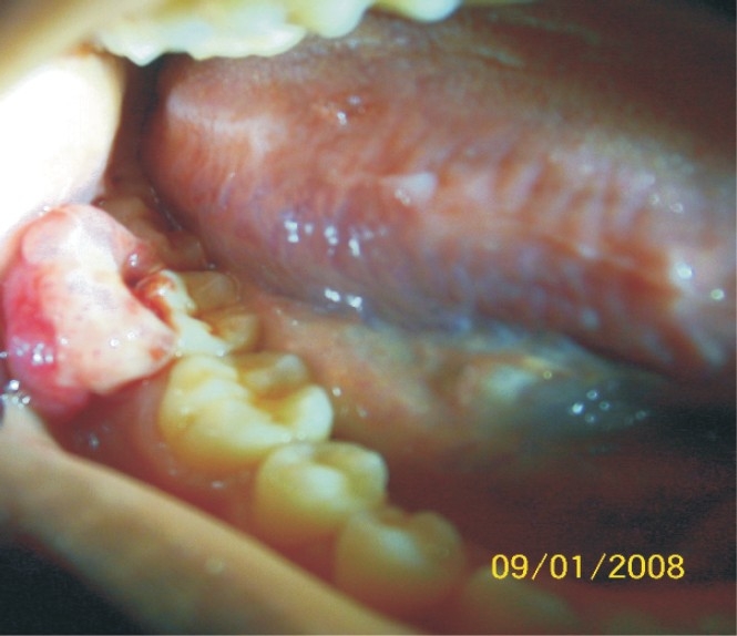
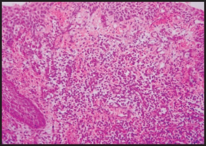
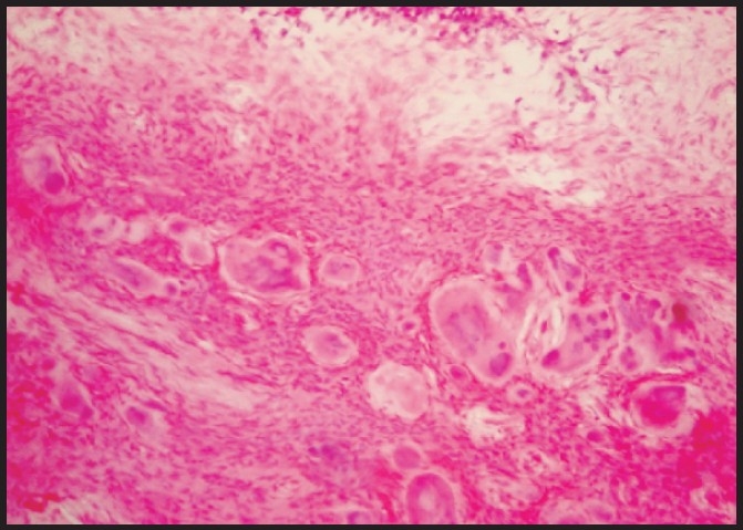
BACKGROUND: The aim of this study was to review the clinicopathologic features of focal reactive gingival lesions at the Lagos University Teaching Hospital, Nigeria. METHODS: A retrospective review of cases of different focal reactive gingival lesions from the records of the Departments of the Oral Biology/Oral Pathology and Oral and Maxillofacial Surgery of the Lagos University Teaching Hospital between 1970 and 2008 was carried out. Available clinical data regarding age, gender, location, estimated duration of the lesion and treatment modality were obtained and analyzed. RESULTS: Prevalence rate of focal reactive gingival lesions was 5.6%. Pyogenic granuloma (PG) was the most common lesions constituting 57% of the cases. Seventeen (9.5%) of the 179 cases of PG were pregnancy induced pyogenic granuloma. The female-to-male ratio was 1.7:1. All the 4 lesions occurred more in female patients than males. The mean age of patients at presentation was 30 ± 16.5 years. The lesions were commonly seen in the second and third decade of life and least commonly seen above the age of 60 years. The lesions were equally distributed on the maxillary and mandibular gingivae, and were mostly located on the buccal gingival of the jaws. Most (51.6%) of the lesions occurred in incisors/canine region. Recurrence of the lesions was seen in 9 cases (2.9%), all pyogenic granuloma. CONCLUSION: Focal reactive gingival lesions are relatively uncommon lesions of the oral cavity with a prevalence rate of 5.6%. The lesions occurred commonly in females, and in third decades of life. Pyogenic granuloma was the most common lesions constituting 57% of all cases.
Figures
References
-
- Scott JH, Symons NBB. 9th edition. London Melbourne and New York: Churchill Livingston Edinburgh; 1982. Introduction to dental anatomy.
-
- Shenoy SS, Dinkar AD. Pyogenic granuloma associated with bone loss in an eight year old child. A case report. J Indian Soc Ped and Prev Dent. 2006;24:201–3. - PubMed
-
- Buchner A, Calderon S, Ramon Y. Localized hyperplastic lesions of the gingival: a clinicopathological study of 302 lesions. J Periodontol. 1977;93:305–9. - PubMed
-
- Stablien MJ, Silverglade LB. Comparative analysis of biopsy specimens from gingival and alveolar mucosa. J. Periodontol. 1985;56(11):671–6. - PubMed
-
- Zarei MR, Chamani G, Amanpoor S. Reactive hyperplasia of the oral cavity in Kerman province, Iran : a review of 172 cases. Br J Oral Maxillofac Surg. 2007;45(4):288–92. - PubMed