Review
. 2011 Oct 3;13(10):1154-60.
doi: 10.1038/ncb2345.

The centrosome cycle: Centriole biogenesis, duplication and inherent asymmetries

Affiliations
Review

The centrosome cycle: Centriole biogenesis, duplication and inherent asymmetries

Erich A Nigg et al. Nat Cell Biol. .

Abstract

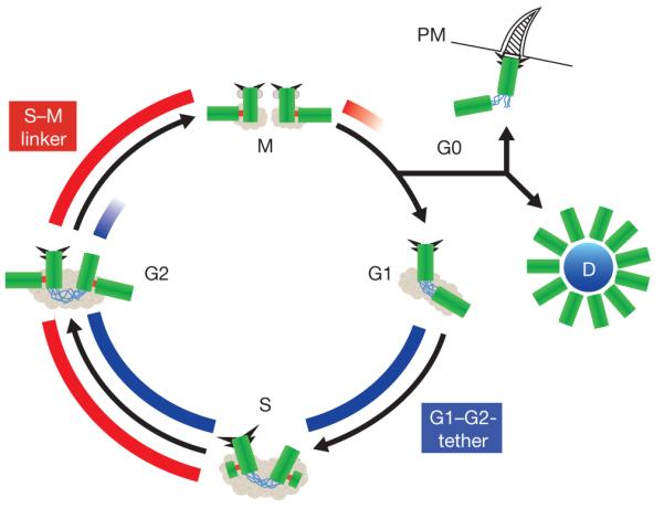
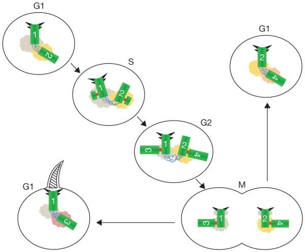
Centrosomes are microtubule-organizing centres of animal cells. They influence the morphology of the microtubule cytoskeleton, function as the base for the primary cilium and serve as a nexus for important signalling pathways. At the core of a typical centrosome are two cylindrical microtubule-based structures termed centrioles, which recruit a matrix of associated pericentriolar material. Cells begin the cell cycle with exactly one centrosome, and the duplication of centrioles is constrained such that it occurs only once per cell cycle and at a specific site in the cell. As a result of this duplication mechanism, the two centrioles differ in age and maturity, and thus have different functions; for example, the older of the two centrioles can initiate the formation of a ciliary axoneme. We discuss spatial aspects of the centrosome duplication cycle, the mechanism of centriole assembly and the possible consequences of the inherent asymmetry of centrioles and centrosomes.


Figures

Figure 1
Figure 1
Centriole biogenesis. This schematic representation of the centriole duplication cycle shows centrioles (green) and PCM (grey), with emphasis on two distinct linker structures. The G1–G2 tether (GGT; blue) connects the proximal ends of the two parental centrioles from G1 to late G2; it is important to ensure microtubule nucleation from a single microtubule organizing centre. The S–M Linker (SML; red) forms during S phase and connects the proximal end of the nascent procentriole to the lateral surface of the mother centriole. The removal of this tight connection in late M phase (disengagement) is an important element of cell cycle control of centriole duplication. Both the molecular components of the GGT and SML as well as the regulation of the formation and dissolution of these structures are expected to be distinct, although some PCM components are likely to be important for both GGT and SML. Also depicted are subdistal and distal appendages (triangles); although readily visible in electron micrographs during interphase, these appendages are difficult to visualize during M phase. In quiescent cells, the appendage-bearing centriole associates with the plasma membrane (PM) and acts as a basal body to form a primary cilium. Finally, in multi-ciliated epithelial cells, multiple centrioles form simultaneously from an amorphous structure termed the deuterosome (D).
Figure 2
Figure 2
Identification of SAS-6 as a key element of the centriolar cartwheel. (a) Immunolocalization of the SAS-6 protein (also known as Bld12p) to the central hub of the cartwheel in Chlamydomonas reinhardtii imaged by electron microscopy. Top images show longitudinal sections through wild-type centrioles; note the immunogold-labelling of the carthwheel by anti-SAS-6 antibodies (right). Bottom; immunogold-labelling of centriole in cross-section, showing that SAS-6 localizes to the central part of the cartwheel (right). Schematic representation (left) shows cartwheel in red. Scale bars,100 nm. Reproduced with permission from ref. 35. (b). Structural model of a SAS-6 oligomer (upper panel) and rotary-metalshadowing electron micrographs of the same structure (lower right panel; schematic representation of structure is shown on the left). Both images emphasize the importance of SAS-6 for conferring ninefold rotational symmetry to the centriole. (Reproduced with permission from ref. 38).
Figure 3
Figure 3
Centriole and centrosome asymmetries. Schematic representation of centrioles (green), with distal and subdistal appendages (triangles), G1–G2 tether (blue), S–M linker (red) and pericentriolar material associated with the base of each centriole. The centrioles are numbered to indicate their origin and age. The centriole marked ‘1’ (centriole 1) is the older of the two centrioles in the G1 cell at the upper left. Centriole 2 formed in the previous cell cycle, as a procentriole adjacent to centriole 1. The centrioles in this cell are disengaged (no S–M linker), but tethered (G1–G2 tether). In S phase new procentrioles grow from each of centrioles 1 and 2 and elongate in G2 phase. These new centrioles (3 and 4) are engaged to their mother centrioles (1 and 2, respectively), but are otherwise equivalent. Centriole 2 acquires appendage proteins at the G2/M transition and appendages proper in the subsequent G1. The two centrosomes segregate at mitosis, with one cell receiving the 1, 3 pair and the other receiving the 2, 4 pair. Although the centriole pairs are morphologically equivalent, there is a functional difference, in that the cell receiving the older mother centriole (centriole 1), is able to form a primary cilium earlier in the cell cycle than the other cell. The pericentriolar material at the base of each centriole is represented in different colours to indicate the possibility that proteins associated with the centrioles might be asymmetrically segregated at mitosis with them.

References

    1. Luders J, Stearns T. Microtubule-organizing centres: a re-evaluation. Nat. Rev. Mol. Cell Biol. 2007;8:161–167. - PubMed
    1. Goetz SC, Anderson KV. The primary cilium: a signalling centre during vertebrate development. Nat. Rev. Genet. 2010;11:331–344. - PMC - PubMed
    1. Carvalho-Santos Z, Azimzadeh J, Pereira-Leal JB, Bettencourt-Dias M. Evolution: Tracing the origins of centrioles, cilia, and flagella. J. Cell Biol. 2011;194:165–175. - PMC - PubMed
    1. Nigg EA, Raff JW. Centrioles, centrosomes, and cilia in health and disease. Cell. 2009;139:663–678. - PubMed
    1. Vaughan S, Dawe HR. Common themes in centriole and centrosome movements. Trends Cell Biol. 2011;21:57–66. - PubMed

Publication types