. 2011 Nov 7;52(12):8646-56.
doi: 10.1167/iovs.11-7570.

Expression and functional roles of caspase-5 in inflammatory responses of human retinal pigment epithelial cells

Affiliations

Expression and functional roles of caspase-5 in inflammatory responses of human retinal pigment epithelial cells

Zong-Mei Bian et al. Invest Ophthalmol Vis Sci. .

Abstract

Purpose: To investigate the expression, activation, and functional involvement of caspase-5 in human retinal pigment epithelial (hRPE) cells.

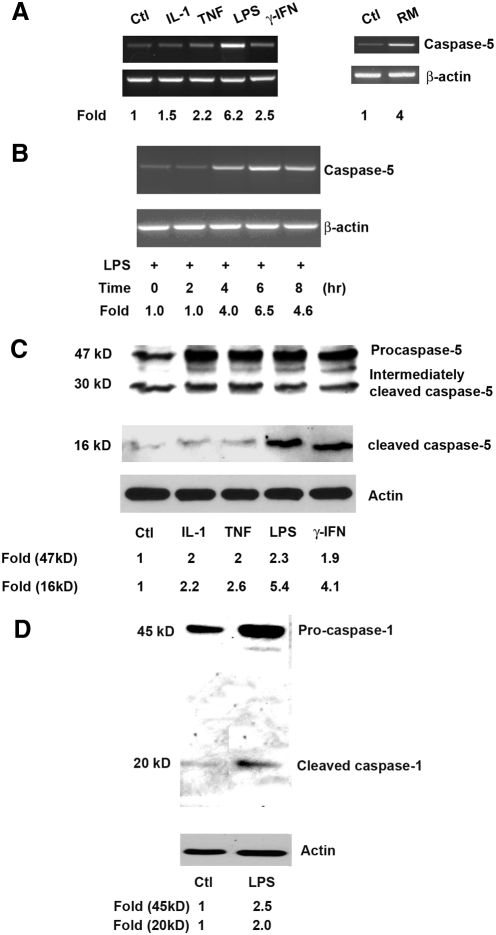
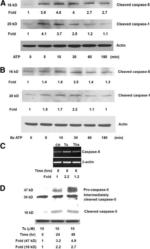
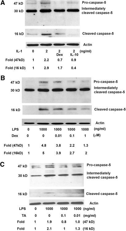
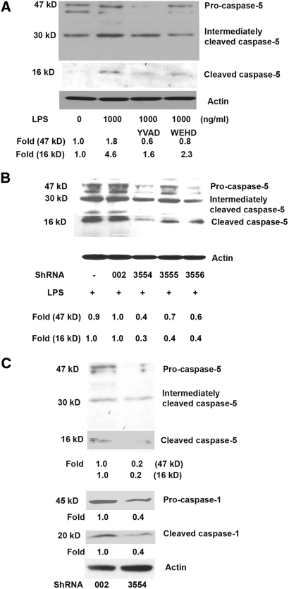
Methods: Expression and activation of caspase-5 in primary cultured hRPE cells, telomerase-immortalized hTERT-RPE1 cells (hTERT-RPE1), or both, were measured after stimulation with proinflammatory agents IL-1β, TNF-α, lipopolysaccharide (LPS), interferon-γ, monocyte coculture, adenosine triphosphate (ATP), or endoplasmic reticulum (ER) stress inducers. Immunomodulating agents dexamethasone (Dex), IL-10, and triamcinolone acetonide (TA) were used to antagonize proinflammatory stimulation. Cell death ELISA and TUNEL staining assays were used to assess apoptosis.

Results: Caspase-5 mRNA expression and protein activation were induced by LPS and monocyte-hRPE coculture. Caspase-5 activation appeared as early as 2 hours after challenge by LPS and consistently increased to 24 hours. Meanwhile, caspase-1 expression and protein activation were induced by LPS. Activation of caspase-5 was blocked or reduced by Dex, IL-10, and TA. Activation of caspase-5 and -1 was also enhanced by ATP and ER stress inducers. Expression and activation of caspase-5 were inhibited by a caspase-1-specific inhibitor. Caspase-5 knockdown reduced caspase-1 protein expression and activation and inhibited TNF-α-induced IL-8 and MCP-1. In contrast to caspase-4, the contribution of caspase-5 to stress-induced apoptosis was moderate.

Conclusions: Caspase-5 mRNA synthesis, protein expression, and catalytic activation were highly regulated in response to various proinflammatory stimuli, ATP, and ER stress inducers. Mutual activation between caspase-5 and -1 suggests caspase-5 may work predominantly in concert with caspase-1 in modulating hRPE inflammatory responses.


Figures

Figure 1.
Figure 1.
Stimulation of caspase-5 mRNA synthesis (A, B) and caspase-5 and -1 protein cleavage (C, D) in hRPE cells. The hRPE cells were cultured either without (untreated; Ctl) or with IL-1β (IL-1, 2 ng/mL), TNF-α (TNF, 20 ng/mL), LPS (1000 ng/mL), IFN-γ (500 U/mL), or overlaid monocytes (RM) and were incubated for 6 hours (A), 0, 2, 4, 6, and 8 hours (B), or 24 hours (C, D). The data shown represent results from a typical experiment. (A, B) The steady state caspase-5 mRNA levels determined by RT-PCR. (C, D) Western blot analysis of caspase-5 protein. The fold changes were calculated by normalization against β-actin and comparison with untreated control (A) or with treated control at 0 hours (B) of mRNA levels in RT-PCR, or by normalization against actin and making a comparison with untreated control levels of caspase-5 in Western blot analysis (C, D). The bands at approximately 30 kDa (C) are presumably either nonspecific bands or intermediately cleaved caspase-5 protein.
Figure 2.
Figure 2.
Induction of caspase-5 catalytic activity by LPS. The hRPE cells were stimulated by LPS (1000 ng/mL, A, B) for 0, 1, 2, 7 (A), and 24 (A, B) hours, with or without caspase -1 inhibitor Z-YVAD-fm (2 μM) or Dex (10−6 M). Two hundred micrograms of protein from the hRPE cell lysate was analyzed in each assay using a caspase-5 assay kit. The data are representative of three independent experiments with similar results, as shown by fold increases in caspase-5 activity. Values represent mean ± SEM. *P < 0.05; ***P < 0.001.
Figure 3.
Figure 3.
Stimulation of human RPE caspase-5 mRNA synthesis (C) and protein maturation (A, B, D) by ATP and ER stress. hRPE cells were pretreated with LPS (1000 ng/mL, A, B) for 4 hours and cultured either with ATP (3 mM) or BzATP (300 μM) for 0, 5, 10, 30, 60, and 100 minutes, tunicamycin (10 μM), or thapsigargin (25 ng/mL) for 6 (C), 0, 24, and 48 hours (D). The data shown represent results from a typical experiment. (C) Steady state caspase-5 mRNA, as determined by RT-PCR. The fold changes were calculated by normalization against β-actin and comparison with control cells without the treatment. (A, B, D) Western blot analysis of caspase-5, caspase-1, and actin proteins. (D) The bands at approximately 30 kDa are presumably either nonspecific bands or intermediately cleaved caspase-5 protein.
Figure 4.
Figure 4.
The effect of Dex, IL-10, and TA on caspase-5 protein production and maturation by IL-1β (A) and LPS (B, C) in hRPE cells. hRPE cells were pretreated with Dex (10−8 or 10−7 to 10−6 M), IL-10 (100 U/mL), or TA (0.1 or 0.01 mg/mL) for 30 minutes and then coincubated with IL-1β (2 ng/mL) or LPS (1000 ng/mL) for an additional 24 hours. Proteins from the whole hRPE cell lysates were detected by anti–caspase-5 antibody specific for pro-caspase-5 and cleaved caspase-5. Fold changes of the cleaved caspase-5 were calculated by relative density between treated and untreated samples, as determined by densitometry after normalization with actin protein.
Figure 5.
Figure 5.
Blockade of caspase-5 activation by caspase-1 inhibitors (A) and caspase-5 and -1 activation by caspase-5 knockdown (B, C). (A) hRPE cells were pretreated with caspase-1 inhibitor Z-YVAD-fmk or caspase-1 and -5 inhibitor Z-WEHD-fmk for 30 minutes and then coincubated with or without LPS (1000 ng/mL) for an additional 24 hours. Proteins from the whole hRPE cell lysates were subjected to Western blot analysis using anti–caspase-5 antibody. (B, C) Whole cell lysates from stably knockdown hTERT-RPE 1 cells with caspase-5 shRNA and control shRNA were detected by antibodies specific for pro-caspase-5 or cleaved caspase-5 or -1. Fold changes of cleaved caspase-5 and -1 were calculated by relative density between scrambled shRNA002 and caspase-5–specific shRNA3554 using densitometry after normalization to actin protein.
Figure 6.
Figure 6.
Inhibition of human RPE IL-8 and MCP-1 production by caspase inhibitors (A, B, E, F) and caspase-5 shRNA knockdown (C, D). hRPE cells (A, B, E, F) were pretreated with caspase-5 and -1 inhibitor Z-WEHD-fmk (2 μM), caspase-1 inhibitor Z-YVAD-fmk (2 μM), or pan-caspase inhibitor Z-VAD-fmk (50 μM) and were costimulated with IL-1β (2 ng/mL), TNF-α (20 ng/mL), or LPS (1000 ng/mL) for 24 hours. (C, D) hTERT-RPE 1 cells were transfected with scrambled shRNA002 or caspase-5–specific shRNA3554. Secretion of IL-8 and MCP-1 was determined by ELISA. Values represent mean ± SEM (n = 3). Statistical analysis was carried out for all values with inhibitor versus the corresponding control without inhibitor (A, B, E, F) or for all values with scrambled shRNA versus caspase-5–specific shRNA. Data are representative of three independent experiments with similar results. Ctrl, untreated; IL-1, IL-1β; TNF, TNF-α.
Figure 7.
Figure 7.
ER stress-induced hRPE apoptotic cell death. hRPE cells were cultured with or without tunicamycin (10 μM) in the presence or absence of caspase-4 inhibitor Z-LEVD-fmk (Z-LEVD, 2 μM) or caspase-5 and -1 inhibitor Z-WEHD-fmk (Z-WEHD, 2 μM) for 48 or 72 hours. Apoptosis was determined by the absorbance difference between A405nm and A490nM using a cell death detection ELISA kit (A). For comparison, tunicamycin-treated cells were assigned as 100% apoptotic cell death (positive control). (B, C) Quantification of the effects of ER stress-induced hRPE cell death by TUNEL assays. (B) TUNEL staining (dark brown), shown at 400× magnification. The hRPE cells were stained by vimentin (red). Left, middle: unstimulated hRPE cells (normal cultures and culture in TUNEL assay); right: hRPE cells treated with tunicamycin showing nuclear condensation and cell shrinkage. (C) Data are expressed as percentage of TUNEL-positive hRPE cells. Values represent mean ± SEM. ***P <0.001, **P < 0.01, and *P < 0.05, compared with tunicamycin treatment without inhibitors. The findings are representative of three independent experiments.

Similar articles

Cited by

References

    1. Munday NA, Vaillancourt JP, Ali A, et al. Molecular cloning and pro-apoptotic activity of ICErelII and ICErelIII, members of the ICE/CED-3 family of cysteine proteases. J Biol Chem. 1995;270:15870–15876 - PubMed
    1. Faucheu C, Blanchet AM, Collard-Dutilleul V, Lalanne JL, Diu-Hercend A. Identification of a cysteine protease closely related to interleukin-1 beta-converting enzyme. Eur J Biochem. 1996;236:207–213 - PubMed
    1. Lin XY, Choi MS, Porter AG. Expression analysis of the human caspase-1 subfamily reveals specific regulation of the CASP5 gene by lipopolysaccharide and interferon-gamma. J Biol Chem. 2000;275:39920–39926 - PubMed
    1. Eckhart L, Kittel C, Gawlas S, et al. Identification of a novel exon encoding the amino-terminus of the predominant caspase-5 variants. Biochem Biophys Res Commun. 2006;348:682–688 - PubMed
    1. Martinon F, Tschopp J. Inflammatory caspases and inflammasomes: master switches of inflammation. Cell Death Differ. 2007;14:10–22 - PubMed

Publication types

MeSH terms