. 2011 Dec 2;286(48):41883-41892.
doi: 10.1074/jbc.M111.301465. Epub 2011 Oct 3.

Histone chaperone FACT coordinates nucleosome interaction through multiple synergistic binding events

Affiliations

Histone chaperone FACT coordinates nucleosome interaction through multiple synergistic binding events

Duane D Winkler et al. J Biol Chem. .

Abstract

In eukaryotic cells, DNA maintenance requires ordered disassembly and re-assembly of chromatin templates. These processes are highly regulated and require extrinsic factors such as chromatin remodelers and histone chaperones. The histone chaperone FACT (facilitates chromatin transcription) is a large heterodimeric complex with roles in transcription, replication, and repair. FACT promotes and subsequently restricts access to DNA as a result of dynamic nucleosome reorganization. However, until now, there lacked a truly quantitative assessment of the critical contacts mediating FACT function. Here, we demonstrate that FACT binds histones, DNA, and intact nucleosomes at nanomolar concentrations. We also determine roles for the histone tails in free histone and nucleosome binding by FACT. Furthermore, we propose that the conserved acidic C-terminal domain of the FACT subunit Spt16 actively displaces nucleosomal DNA to provide access to the histone octamer. Experiments with tri-nucleosome arrays indicate a possible mode for FACT binding within chromatin. Together, the data reveal that specific FACT subunits synchronize interactions with various target sites on individual nucleosomes to generate a high affinity binding event and promote reorganization.


Figures

FIGURE 1.
FIGURE 1.
FACT preferentially binds H2A-H2B over H3-H4, yet both interactions are enhanced by the histone tails. A shows the normalized fluorescence change upon titration of FACT (log [FACT]) into fluorescently labeled H2A-H2B and TL H2A-H2B. Fluorescence change (F.C.) occurs as a result of direct protein/protein interaction. A 7-fold decrease in affinity between FACT and H2A-H2B is observed when these basic extensions are removed from the histones (binding curve shifts right to higher FACT concentrations). B displays the binding curves for FACT with H3-H4 and TL H3-H4. FACT·H3-H4 binding occurs at high nanomolar concentrations, yet deletion of the N- and C-terminal tails pushes binding out of the nanomolar range. The error bars represent the standard error within individual data points. The total data points (N) for a single experiment are 12.
FIGURE 2.
FIGURE 2.
Spt16 subunit and its acidic C-terminal domain coordinate high affinity FACT interaction(s) with H2A-H2B. A identifies the FACT subunit (Spt16 or SSRP1) responsible for the interaction with H2A-H2B. The lack of discernible fluorescence change for the SSRP1·H2A-H2B titration indicates a lack of binding within this concentration range. The Spt16 subunit binds H2A-H2B with slightly lower affinity than full-length FACT. The FACT·H2A-H2B binding curve from Fig. 1 is displayed as a dashed line to provide a clear comparison. B shows the binding curve for an Spt16 CTD-deleted FACT construct (FACTΔCTD) and H2A-H2B. Without the Spt16 CTD, the binding affinity for H2A-H2B decreases ∼6-fold. The error bars represent the standard error within individual data points. The total data points (N) for a single experiment are 12.
FIGURE 3.
FIGURE 3.
FACT competes with DNA for a shared interaction interface on H2A-H2B but cannot compete H3-H4 from DNA. Histones can readily bind DNA at physiological salt concentrations in vitro, as seen by native PAGE. The use of fluorescently labeled histones allows visualization by two distinct means (UV, top panels, to monitor DNA; and fluorescence scanning, bottom panels, to monitor histones). The H2A-H2B dimer forms many stable complexes with a 207-bp 601 sequence DNA construct (A). Incubation with H3-H4 will form a stable tetrasome where DNA is wrapped around a single (H3-H4)2 tetramer (B). The appearance of accumulating free DNA (top left panel, lanes 4–8) and a higher order band running similar to a preformed FACT·H2A-H2B complex (bottom left panel, lanes 6–8) confirms that FACT can compete H2A-H2B from DNA but cannot form a ternary complex with DNA-bound H2A-H2B. Right panels show that, in contrast, FACT cannot remove H3-H4 from DNA once bound. No evidence for free DNA, a FACT·H3-H4 complex, or ternary complex is observed upon FACT titration (B, lanes 4–8). Red asterisks are displayed next to the furthest running histone·DNA species to aid specific band comparisons between the UV and fluorescence gel images.
FIGURE 4.
FIGURE 4.
Linker DNA is required for high affinity interaction between SSRP1 and nucleosomes. Nuc147 lacks the symmetrical 30-bp linker DNA extensions of Nuc207. FACT and Spt16 can bind either form with high affinity (Kd(app) <100 nm). However, as seen in A, the SSRP1 subunit cannot bind the 147-bp nucleosome in the nanomolar range. However, SSRP1 binds Nuc207 with high affinity (B), which suggests that linker DNA is a target of SSRP1 binding. B also shows that both Spt16 and SSRP1 subunits together in complex are essential for achieving the highest affinity binding event with Nuc207. The error bars represent the standard error within individual data points. The total data points (N) for a single experiment are 12.
FIGURE 5.
FIGURE 5.
Stoichiometry and positions of FACT within tri-nucleosome arrays. A–D show stoichiometry measurements for FACT with the Nuc207, Nuc147, and two distinct tri-nucleosome constructs. The tri-nucleosomes are labeled LE (linker ended), which contain 30-bp linker arms extending from the terminal nucleosomes, and NLE (non-linker ended), which lacks terminal linkers. To measure stoichiometries, the labeled probe ((tri)nucleosomes) was kept constant 5–10-fold over the measured Kd(app) so that any addition of FACT results in direct interaction. The fluorescence change deviates from linearity at the point of saturation. The presence of terminal linker DNA on the (tri)nucleosome (A and C) increases the stoichiometry compared with the Nuc147 and NLE tri-nucleosome. The error bars represent the standard error within individual data points. The total data points (N) for a single experiment are 12.
FIGURE 6.
FIGURE 6.
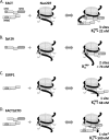
Multiple interactions between FACT and nucleosome produce a singular high affinity binding event. The critical nucleosome-binding sites on FACT, as identified in our study, are shaded dark and labeled as CTD, N-terminal domain (NTD), and HMG for the acidic C-terminal domain of Spt16, N-terminal domain of Spt16, and the HMG-1 DNA binding site on SSRP1, respectively. A illustrates the inclusion of the three binding sites in the modulation of an intact nucleosome by full-length FACT. The Spt16 N-terminal domain binds histone tails, and the CTD displaces DNA for binding by the SSRP1 HMG-1 domain. Elimination of the SSRP1 subunit reduces the number of interaction sites to two and thus decreases the overall affinity for Nuc207 (B). The SSRP1 subunit alone, in C, can only promote a single high affinity interaction with Nuc207 through accessible linker DNA. Deletion of the Spt16 CTD from FACT not only eliminates a direct interaction interface with the core histones but seemingly blocks DNA binding by the SSRP1 HMG-1 domain. The overall affinity for Nuc207 decreases ∼20-fold.

References

    1. Luger K., Mäder A. W., Richmond R. K., Sargent D. F., Richmond T. J. (1997) Nature 389, 251–260 - PubMed
    1. Oohara I., Wada A. (1987) J. Mol. Biol. 196, 399–411 - PubMed
    1. Wilhelm F. X., Wilhelm M. L., Erard M., Duane M. P. (1978) Nucleic Acids Residues 5, 505–521 - PMC - PubMed
    1. Li Q., Burgess R., Zhang Z. (2011) Biochim. Biophys. Acta, in press - PMC - PubMed
    1. Tremethick D. J. (2007) Cell 128, 651–654 - PubMed

Publication types

MeSH terms

LinkOut - more resources