Review
. 2011 Oct 3;18(10):666-77.
doi: 10.1101/lm.2023011. Print 2011.

Neural circuitry and plasticity mechanisms underlying delay eyeblink conditioning

Affiliations
Review

Neural circuitry and plasticity mechanisms underlying delay eyeblink conditioning

John H Freeman et al. Learn Mem. .

Abstract

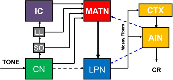
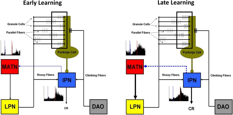
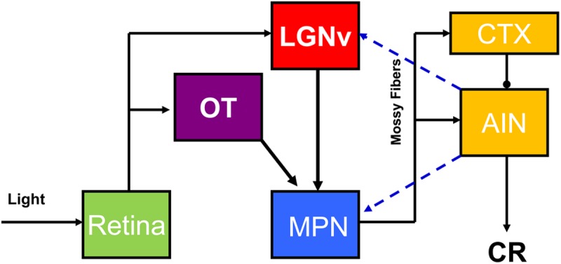
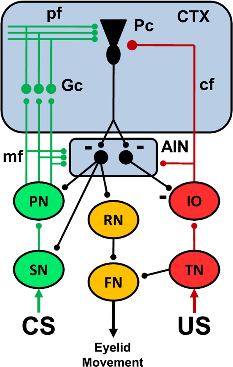
Pavlovian eyeblink conditioning has been used extensively as a model system for examining the neural mechanisms underlying associative learning. Delay eyeblink conditioning depends on the intermediate cerebellum ipsilateral to the conditioned eye. Evidence favors a two-site plasticity model within the cerebellum with long-term depression of parallel fiber synapses on Purkinje cells and long-term potentiation of mossy fiber synapses on neurons in the anterior interpositus nucleus. Conditioned stimulus and unconditioned stimulus inputs arise from the pontine nuclei and inferior olive, respectively, converging in the cerebellar cortex and deep nuclei. Projections from subcortical sensory nuclei to the pontine nuclei that are necessary for eyeblink conditioning are beginning to be identified, and recent studies indicate that there are dynamic interactions between sensory thalamic nuclei and the cerebellum during eyeblink conditioning. Cerebellar output is projected to the magnocellular red nucleus and then to the motor nuclei that generate the blink response(s). Tremendous progress has been made toward determining the neural mechanisms of delay eyeblink conditioning but there are still significant gaps in our understanding of the necessary neural circuitry and plasticity mechanisms underlying cerebellar learning.


Figures

Figure 1.
Figure 1.
Examples of single unit activity (spikes/sec) recorded from the anterior interpositus nucleus and corresponding average eyelid EMG activity in well-trained rats during CS-US trials. The green line represents CS onset and the red line represents US onset. Bars in the histograms are 10 msec. Note the increase in activity after onset of the CS.
Figure 2.
Figure 2.
Hypothesized auditory CS pathway for eyeblink conditioning. There are parallel inputs into the medial auditory thalamic nuclei (MATN) from the cochlear nucleus (CN), superior olive (SO), nucleus of the lateral lemniscus (LL), and inferior colliculus (IC). Auditory input is then projected from the MATN to the lateral pontine nuclei (LPN), which project to the cerebellar cortex (CTX) and anterior interpositus nucleus (AIN). Hypothesized feedback projections from the AIN to the LPN and MATN are also shown (blue dashed lines).
Figure 3.
Figure 3.
Hypothesized role of medial auditory thalamic plasticity in cerebellar learning. Paired stimulus inputs from the lateral pontine nuclei (LPN) and dorsal accessory inferior olive (DAO) converge on Purkinje cells in the cerebellar cortex during the early stages of eyeblink conditioning (left). Learning is initiated by CS-activated parallel fibers that arrive at Purkinje cells nearly simultaneously with climbing fiber input (window). No learning-related plasticity is evident in the interpositus nucleus (IPN) or medial auditory thalamic nuclei (MATN) during the initial training trials. Paired stimulus inputs from the LPN and DAO continue to converge on Purkinje cells after learning starts to emerge (right). Learning is potentiated by an increase (relative to initial training) in parallel fiber activity that occurs nearly simultaneously with climbing fiber input (window) and mossy fiber activity in the IPN. Clear learning-related plasticity is evident in IPN and MATN during this later stage of learning. An increase in learning-related excitatory cerebellar feedback to the MATN (blue dotted line) may, in turn, increase the MATN output to the LPN and corresponding mossy fiber projection into the cerebellum to further facilitate learning.
Figure 4.
Figure 4.
Hypothesized visual CS pathway necessary for delay eyeblink conditioning. Inputs from the retina to the ventral lateral geniculate (LGNv) and nucleus of the optic tract (OT) are relayed in parallel to the cerebellar cortex (CTX) and anterior interpositus nucleus (AIN) via the medial pontine nuclei (MPN). Hypothesized feedback projections from the AIN to the MPN and LGNv are also shown (blue dashed lines).
Figure 5.
Figure 5.
A simplified schematic diagram of the neural circuitry underlying eyeblink conditioning. The cerebellar anterior interpositus nucleus (AIN) and Purkinje cells (Pc) in the cerebellar cortex (CTX) receive convergent input from the conditioned stimulus (CS, green) and unconditioned stimulus (US, red) neural pathways. The pathways for different CSs include subcortical sensory nuclei (SN), basilar pontine nuclei (PN), mossy fiber (mf) projection to the AIN and cortical granule cells (Gc), and the parallel fiber (pf) projection to Purkinje cells. The US pathway includes the trigeminal nucleus (TN), dorsal accessory division of the inferior olive (IO), and the climbing fiber (cf) projection to the AIN and Pc. The output pathway for performance of the conditioned response (orange) includes the AIN projection to the red nucleus (RN) and its projection to facial motor nucleus (FN) which causes eyelid closure. The unconditioned response is elicited by activation of the TN, which then activates the FN. Feedback projections from the AIN to the PN, SN, and IO regulate CS and US input to facilitate acquisition and maintain plasticity within the cerebellum. Inhibitory synapses are depicted by a minus sign. All other synapses are excitatory.

References

    1. Aiba A, Kano M, Chen C, Stanton ME, Fox GD, Herrup K, Zwingman TA, Tonegawa S 1994. Deficient cerebellar long-term depression and impaired motor learning in mGluR1 mutant mice. Cell 79: 377–388 - PubMed
    1. Aksenov D, Serdyukova N, Irwin K, Bracha V 2004. GABA neurotransmission in the cerebellar interposed nuclei: Involvement in classically conditioned eyeblinks and neuronal activity. J Neurophysiol 91: 719–727 - PubMed
    1. Albus JS 1971. A theory of cerebellar function. Math Biosci 10: 25–61
    1. Allen MT, Padilla Y, Gluck MA 2002. Ibotenic acid lesions of the medial septum retard delay eyeblink conditioning in rabbits (Oryctolagus cuniculus). Behav Neurosci 116: 733–738 - PubMed
    1. Andersson G, Garwicz M, Hesslow G 1988. Evidence for a GABA-mediated cerebellar inhibition of the inferior olive in the cat. Exp Brain Res 72: 450–456 - PubMed

LinkOut - more resources