. 2011 Oct 18;108(42):17314-9.
doi: 10.1073/pnas.1110402108. Epub 2011 Oct 3.

Crystal structure of the octameric pore of staphylococcal γ-hemolysin reveals the β-barrel pore formation mechanism by two components

Affiliations

Crystal structure of the octameric pore of staphylococcal γ-hemolysin reveals the β-barrel pore formation mechanism by two components

Keitaro Yamashita et al. Proc Natl Acad Sci U S A. .

Abstract

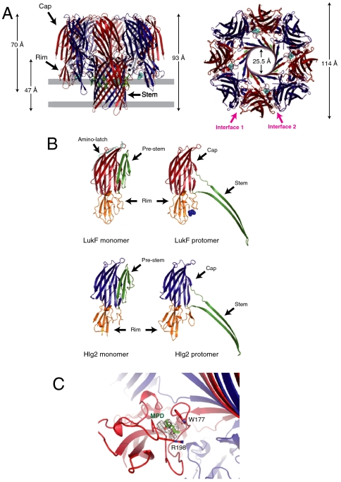
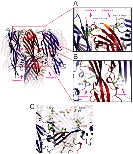
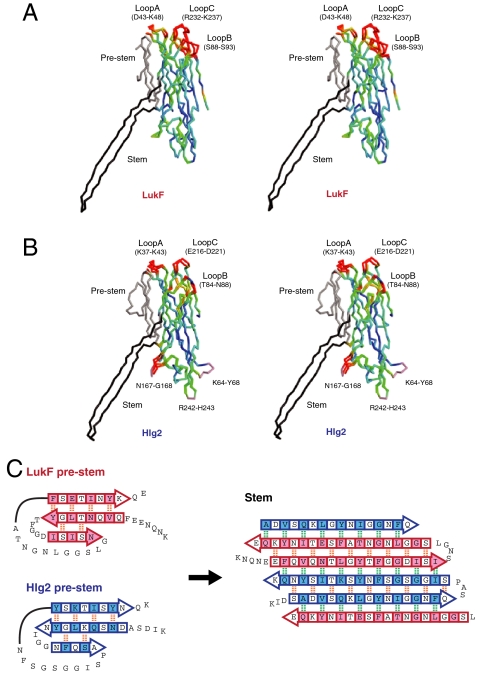
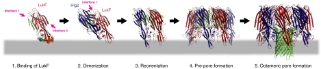
Staphylococcal γ-hemolysin is a bicomponent pore-forming toxin composed of LukF and Hlg2. These proteins are expressed as water-soluble monomers and then assemble into the oligomeric pore form on the target cell. Here, we report the crystal structure of the octameric pore form of γ-hemolysin at 2.5 Å resolution, which is the first high-resolution structure of a β-barrel transmembrane protein composed of two proteins reported to date. The octameric assembly consists of four molecules of LukF and Hlg2 located alternately in a circular pattern, which explains the biochemical data accumulated over the past two decades. The structure, in combination with the monomeric forms, demonstrates the elaborate molecular machinery involved in pore formation by two different molecules, in which interprotomer electrostatic interactions using loops connecting β2 and β3 (loop A: Asp43-Lys48 of LukF and Lys37-Lys43 of Hlg2) play pivotal roles as the structural determinants for assembly through unwinding of the N-terminal β-strands (amino-latch) of the adjacent protomer, releasing the transmembrane stem domain folded into a β-sheet in the monomer (prestem), and interaction with the adjacent protomer.


Conflict of interest statement

The authors declare no conflict of interest.

Figures

Fig. 1.
Fig. 1.
Overall octameric pore structure of γHL. (A) Side and top views of the heptamer. LukF and Hlg2 are shown in red and blue, respectively. MPD molecules bound with LukF are shown as cyan spheres. The aromatic side chains located around the putative membrane surface are shown as green sticks. The putative membrane region is also shown in gray. (B) Structures of the protomers of LukF (Upper) and Hlg2 (Lower). Monomeric structures of each molecule are also shown. Red, cap of LukF; blue, cap of Hlg2; green, stem (prestem in monomers); orange, rim; cyan, amino-latch. Blue spheres represent MPD bound with LukF protomer. (C) Close-up view of the MPD binding site. The bound MPD, Trp177, and Arg198 are shown as sticks. The Fo-Fc map (contoured at 1.5σ) around the MPD is also shown.
Fig. 2.
Fig. 2.
Electrostatic interactions at the interprotomer interface. (A) Interactions between loop A and β-strand 1. The N-terminal residues that are not necessary for pore formation are shown in orange. (B) Electrostatic interaction between Arg219 (LukF) and Glu145 (Hlg2) located at interface 2. Structurally corresponding residues at interface 1 [Asp194 (Hlg2) and Arg151 (LukF)] are also shown. (C) Ion pairs stabilizing the upper end of the β-barrel.
Fig. 3.
Fig. 3.
Structure comparison between monomer and protomer. (A and B) Stereo representations of the superposed monomer and protomer LukF (A) and Hlg2 (B). The Cα trace of each molecule is colored according to the distance between Cα atoms of corresponding residues in monomer and protomer from blue (0 Å) to red (1.5 Å). Residues with Cα distances greater than 1.5 Å are shown in red. The prestem and stem are shown in gray and black, respectively. Loops disordered in the monomer are also shown in pink. (C) Schematic representation of the structural change between prestem (Left) and stem. Arrows represent β-strands. Intra- and interprotomer backbone hydrogen bonds are shown as orange and green dashed lines, respectively. Residues the side chains of which face the hydrophobic environment (interface between prestem and cap domain in monomers or outside of the β-barrel in octamer) are shown in pink (LukF) and/or blue (Hlg2) shaded boxes.
Fig. 4.
Fig. 4.
Mechanism of assembly for staphylococcal γ-HL. (1) Binding of LukF. LukF binds to the erythrocyte surface via Tyr72, Trp257, Phe260, and Tyr 261 (shown as orange spheres) inclined with respect to its molecular axis. Trp177 and Arg198 (shown as green spheres) capture lipid head groups. As a consequence of the orientation, interface 1 is exposed to the surface, whereas interface 2 is hindered. The membrane is shown as a gray bar. (2) Dimerization. Hlg2 binds to LukF through the surface-exposed interface 1. Upon binding, the amino-latch of Hlg2 is released from the β-sheet due to the structural clash with loop A (LukF). At this step, the prestem is not released completely from the cap domain. (3) Reorientation. The heterodimer changes the orientation to expose interface 2. Binding of Hlg2 with the proteinaceous component(s) may induce this motion. (4) Prepore formation. The heterodimer assembles into a prepore. Upon octameric assembly, the long amino-latch of LukF [yellow in (3)] is released from the cap domain. The electrostatic interactions between loop A and β-1 are formed completely in both interfaces, which induces a conformation change of the prestem into the partially unfolded state and release of the prestem from the cap domain (green loops). (5) Octameric pore formation. The prestem completely unfolds and all hydrogen bonds are disrupted. Unfolded prepore penetrates into the membrane and forms a transmembrane β-barrel (green cartoon).

References

    1. Iacovache I, van der Goot FG, Pernot L. Pore formation: An ancient yet complex form of attack. Biochim Biophys Acta. 2008;1778:1611–1623. - PubMed
    1. Peitsch MC, Tschopp J. Assembly of macromolecular pores by immune defense systems. Curr Opin Cell Biol. 1991;3:710–716. - PubMed
    1. Gouaux E. Channel-forming toxins: tales of transformation. Curr Opin Struct Biol. 1997;7:566–573. - PubMed
    1. Parker MW, Feil SC. Pore-forming protein toxins: From structure to function. Prog Biophys Mol Biol. 2005;88:91–142. - PubMed
    1. Menestrina G, Serra MD, Prevost G. Mode of action of beta-barrel pore-forming toxins of the staphylococcal alpha-hemolysin family. Toxicon. 2001;39:1661–1672. - PubMed

Publication types

MeSH terms

Associated data

LinkOut - more resources