. 2011 Nov 8;108(45):18260-5.
doi: 10.1073/pnas.1108228108. Epub 2011 Oct 3.

Molecular hijacking of siroheme for the synthesis of heme and d1 heme

Affiliations

Molecular hijacking of siroheme for the synthesis of heme and d1 heme

Shilpa Bali et al. Proc Natl Acad Sci U S A. .

Abstract

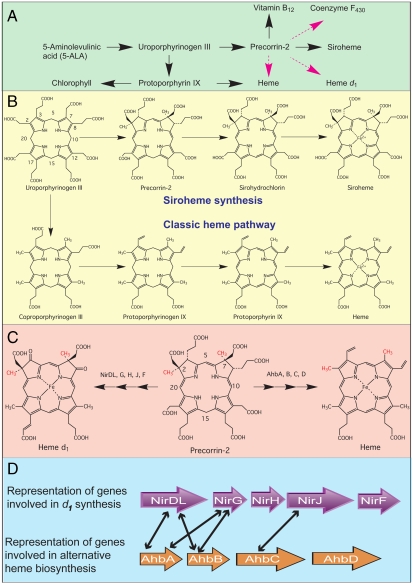
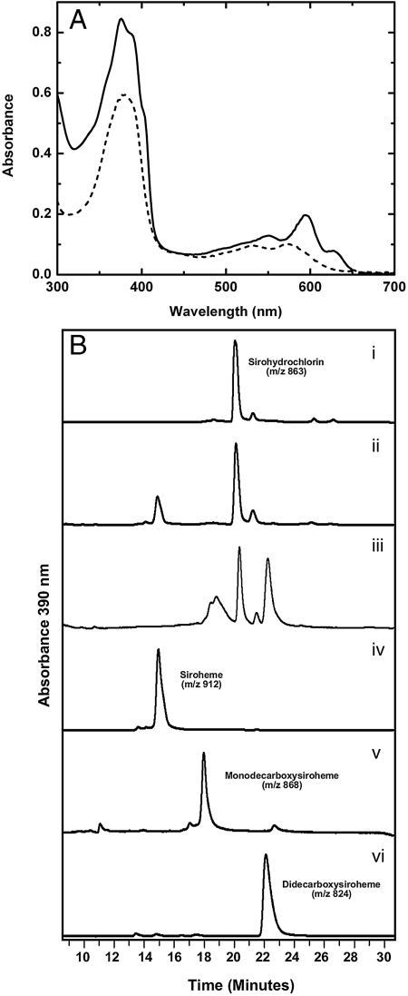
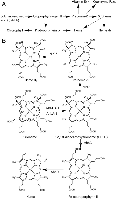
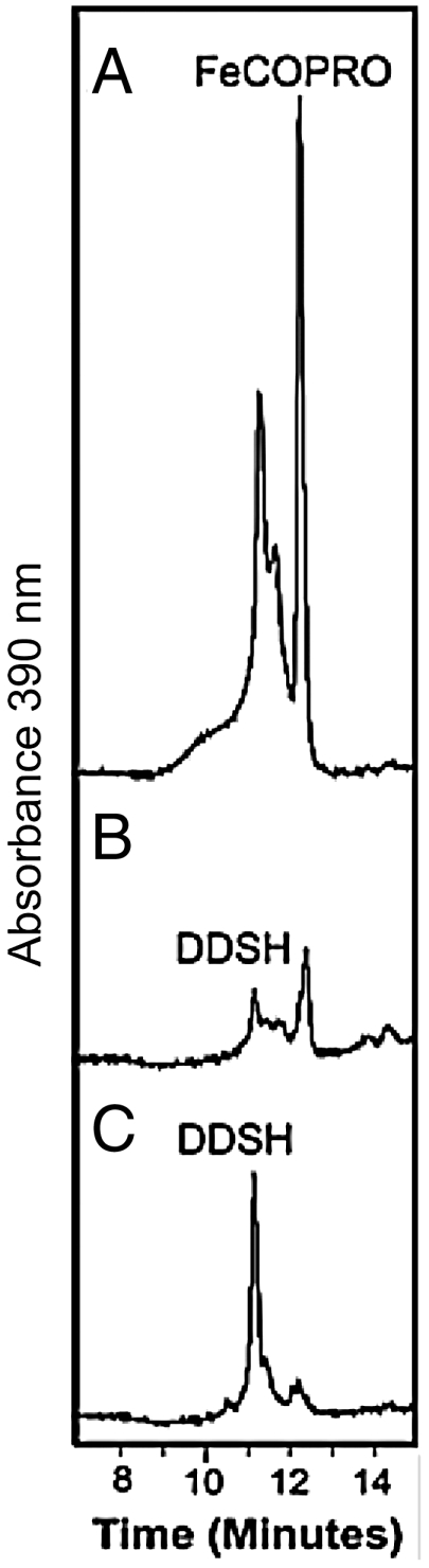
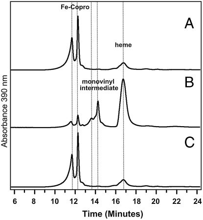
Modified tetrapyrroles such as chlorophyll, heme, siroheme, vitamin B(12), coenzyme F(430), and heme d(1) underpin a wide range of essential biological functions in all domains of life, and it is therefore surprising that the syntheses of many of these life pigments remain poorly understood. It is known that the construction of the central molecular framework of modified tetrapyrroles is mediated via a common, core pathway. Herein a further branch of the modified tetrapyrrole biosynthesis pathway is described in denitrifying and sulfate-reducing bacteria as well as the Archaea. This process entails the hijacking of siroheme, the prosthetic group of sulfite and nitrite reductase, and its processing into heme and d(1) heme. The initial step in these transformations involves the decarboxylation of siroheme to give didecarboxysiroheme. For d(1) heme synthesis this intermediate has to undergo the replacement of two propionate side chains with oxygen functionalities and the introduction of a double bond into a further peripheral side chain. For heme synthesis didecarboxysiroheme is converted into Fe-coproporphyrin by oxidative loss of two acetic acid side chains. Fe-coproporphyrin is then transformed into heme by the oxidative decarboxylation of two propionate side chains. The mechanisms of these reactions are discussed and the evolutionary significance of another role for siroheme is examined.


Conflict of interest statement

The authors declare no conflict of interest.

Figures

Fig. 1.
Fig. 1.
Pathways and genes of modified tetrapyrrole biosynthesis. (A) Outline syntheses of modified tetrapyrroles highlighting key intermediates along the branched pathway. Known reaction sequences are shown in black whereas those not yet elucidated are highlighted in magenta. (B) The known transformation of uroporphyrinogen III into siroheme and heme is shown. The numbering system for the tetrapyrrole macrocycle is highlighted on uroporphyrinogen III. (C) Precorrin-2 as the template for both d1 heme and heme synthesis is shown, with the latter derived via the alternative heme pathway. In both cases the two methyl groups at C2 and C7, highlighted in red, are derived from AdoMet. The enzymes involved in the transformation of precorrin-2 into d1 heme (Nir enzymes) and heme (Ahb enzymes) are shown. (D) The similarity between some of the Nir proteins implicated in d1 synthesis and proteins found in sulfate-reducing bacteria and Archaea thought to be associated with the Ahb pathway are indicated by black arrows.
Fig. 2.
Fig. 2.
Siroheme decarboxylase activity of Nir proteins. (A) UV-visible absorption spectra of sirohydrochlorin (solid line) and didecarboxysiroheme (dashed line), under anaerobic conditions in 50 mM potassium phosphate buffer at pH 8. (B) HPLC traces of tetrapyrrole derivatives observed after incubation of sirohydrochlorin (iiii), and incubation of siroheme (ivvi), with the absorbance recorded at 390 nm. (B, i) HPLC trace of sirohydrochlorin. (B, ii) HPLC trace of reaction containing E. coli cell lysate harboring empty expression vector and sirohydrochlorin, note the new compound at 15 min. (B, iii) HPLC trace of reaction containing NirD-LGH and sirohydrochlorin. (B, iv) HPLC trace of siroheme. (B, v) HPLC trace of reaction containing NirE,D-L and siroheme. (B, vi) HPLC trace of reaction containing NirD-LGH and siroheme. Siroheme was used at a final concentration of 50 µM in each assay.
Fig. 3.
Fig. 3.
Deduced pathway from siroheme to d1 and heme. (A) The corrected modified tetrapyrrole pathway is shown with siroheme now acting as an intermediate for heme and d1 heme synthesis. This pathway can be compared to Fig. 1C. (B) The branched pathway from siroheme to d1 and heme is shown together with the enzymes that are thought to be involved with each particular step. Didecarboxysiroheme is generated from siroheme by the sequential decarboxylation of the side chains attached to C12 and C18. For d1 synthesis, didecarboxysiroheme is modified by replacement of the propionate side chains at C3 and C8 with oxo groups in a reaction likely catalyzed by NirJ to generate a pre-d1 intermediate, which is converted into d1 by the introduction of a double bond on the propionate side chain on C17. For heme synthesis, didecarboxysiroheme is modified by the action of AhbC to remove the acetic acid side chains attached to C2 and C7 to give Fe-coproporphyrin. This intermediate is converted into heme by AhbD through the oxidative decarboxylation of propionate side chains on C3 and C8.
Fig. 4.
Fig. 4.
M. barkeri AhbC catalyzes conversion of didicarboxysiroheme into Fe-coproporphyrin III. HPLC traces of the tetrapyrrole derivates observed at 390 nm after anaerobic incubations of (A) E. coli cell-free extracts overexpressing M. barkeri AhbC with didicarboxysiroheme (DDSH), AdoMet and the reducing agent sodium dithionite, (B) E. coli cell-free extracts overexpressing M. barkeri AhbC with DDSH and sodium dithionite, (C) E. coli cell-free extracts with DDSH, AdoMet, and sodium dithionite. AdoMet and sodium dithionite were used at final concentrations of 500 µM and 8.5 mM, respectively. Note that Fe-coproporphyrin III (Fe-Copro) is eluted in two peaks from the HPLC column; both species have the same m/z value of 708 by MS.
Fig. 5.
Fig. 5.
AhbD catalyzed Fe-coproporphyrin oxidase activity. HPLC traces of observed porphyrin derivatives at 390 nm in reaction containing, (A) Fe-coproporphyrin, cofactor mix, and cell-free extract of E. coli harboring empty expression vector, (B) Fe-coproporphyrin, cofactor mix, and cell-free extract of E. coli overexpressing D. desulfuricans AhbD, and (C) Fe-coproporphyrin, cell-free extract containing D. desulfuricans AhbD without AdoMet in the cofactor mix. Cofactor mix consisted of 500 µM AdoMet and NADPH with 0.3% (vol/vol) Triton X-100 in each assay. Fe-coproporphyrin was used at a final concentration of 25 µM, and is eluted as two close peaks from the HPLC column but with same m/z value of 708 by MS.

References

    1. Horowitz NH. On the evolution of biochemical syntheses. Proc Natl Acad Sci USA. 1945;31:153–157. - PMC - PubMed
    1. Jensen RA. Enzyme recruitment in evolution of new function. Annu Rev Microbiol. 1976;30:409–425. - PubMed
    1. Holliday GL, et al. Evolution of enzymes and pathways for the biosynthesis of cofactors. Nat Prod Rep. 2007;24:972–987. - PubMed
    1. Layer G, Reichelt J, Jahn D, Heinz DW. Structure and function of enzymes in heme biosynthesis. Protein Sci. 2010;19:1137–1161. - PMC - PubMed
    1. Warren MJ, Raux E, Schubert HL, Escalante-Semerena JC. The biosynthesis of adenosylcobalamin vitamin B12. Nat Prod Rep. 2002;19:390–412. - PubMed

Publication types

LinkOut - more resources