. 2011 Oct 11;108(41):17111-6.
doi: 10.1073/pnas.1108121108. Epub 2011 Oct 3.

Chronic inflammation promotes myeloid-derived suppressor cell activation blocking antitumor immunity in transgenic mouse melanoma model

Affiliations

Chronic inflammation promotes myeloid-derived suppressor cell activation blocking antitumor immunity in transgenic mouse melanoma model

Christiane Meyer et al. Proc Natl Acad Sci U S A. .

Abstract

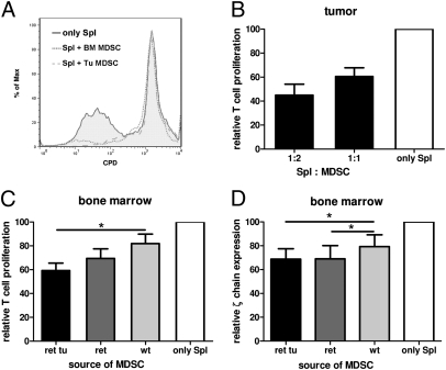
Tumor microenvironment is characterized by chronic inflammation represented by infiltrating leukocytes and soluble mediators, which lead to a local and systemic immunosuppression associated with cancer progression. Here, we used the ret transgenic spontaneous murine melanoma model that mimics human melanoma. Skin tumors and metastatic lymph nodes showed increased levels of inflammatory factors such as IL-1β, GM-CSF, and IFN-γ, which correlated with tumor progression. Moreover, Gr1(+)CD11b(+) myeloid-derived suppressor cells (MDSCs), known to inhibit tumor reactive T cells, were enriched in melanoma lesions and lymphatic organs during tumor progression. MDSC infiltration was associated with a strong TCR ζ-chain down-regulation in all T cells. Coculturing normal splenocytes with tumor-derived MDSC induced a decreased T-cell proliferation and ζ-chain expression, verifying the MDSC immunosuppressive function and suggesting that the tumor inflammatory microenvironment supports MDSC recruitment and immunosuppressive activity. Indeed, upon manipulation of the melanoma microenvironment with the phosphodiesterase-5 inhibitor sildenafil, we observed reduced levels of numerous inflammatory mediators (e.g., IL-1β, IL-6, VEGF, S100A9) in association with decreased MDSC amounts and immunosuppressive function, indicating an antiinflammatory effect of sildenafil. This led to a partial restoration of ζ-chain expression in T cells and to a significantly increased survival of tumor-bearing mice. CD8 T-cell depletion resulted in an abrogation of sildenafil beneficial outcome, suggesting the involvement of MDSC and CD8 T cells in the observed therapeutic effects. Our data imply that inhibition of chronic inflammation in the tumor microenvironment should be applied in conjunction with melanoma immunotherapies to increase their efficacy.


Conflict of interest statement

The authors declare no conflict of interest.

Figures

Fig. 1.
Fig. 1.
Inflammatory mediators in tumor lesions of transgenic mice. IFN-γ, IL-1β, and GM-CSF were measured in primary tumors (AC) and metastatic LN (DF) by Bio-Plex and plotted against the duration of tumor growth between first visible tumor signs and mouse death. Each group included seven to 12 mice. The correlation between two variables was calculated by using a linear regression analysis.
Fig. 2.
Fig. 2.
Analysis of MDSCs and TCR ζ-chain in tumors and lymphatic organs of transgenic mice. Cells from mice with (ret tu) or without (ret) macroscopic tumors and nontransgenic littermates (wt) were assessed by flow cytometry. (A) A representative dot plot of primary tumor is shown. (B) The weight of each tumor sample was plotted against the percentage of tumor-infiltrating Gr1+CD11b+ MDSCs within CD45.2+ leukocytes. (C) The duration of tumor progression was plotted against the percentage of MDSCs among live cells. The correlation was calculated by a linear regression analysis. (D) Cumulative data for MDSCs in metastatic LNs are expressed as the percentage of leukocytes (mean and SE from 10–31 mice). (E) ζ-Chain level in CD3+CD4+ TILs expressed as mean florescence intensity (MFI) and plotted against the tumor weight (n = 12). The correlation was analyzed by using a linear regression analysis. (F) Cumulative data for ζ-chain expression in metastatic LNs are shown as MFI (mean and SE; n = 5–12 mice per group; *P < 0.05).
Fig. 3.
Fig. 3.
Immunosuppressive activity of MDSCs from transgenic mice. (AC) MDSCs were isolated from skin tumors and BM, cocultured with C57BL/6 splenocytes labeled with CPD eFluor 670, and stimulated with anti-CD3 and anti-CD28 mAbs. (A) A histogram from one representative experiment of four is shown. Cumulative data (mean and SE; n = 4–6 mice per group) for the activity of MDSCs isolated from tumors (B) or BM (C) are shown as a ratio between T-cell proliferation levels with and without MDSCs. The proliferation of splenocytes alone (only Spl) was considered as 100%. Spl:MDSC ratios were as indicated (B) or were 1:1 (C). (D) FACS-sorted MDSCs were coincubated with nonstimulated C57BL/6 splenocytes at the Spl:MDSC ratio of 0.6:1 followed by the ζ-chain detection. Results are depicted as a ratio between ζ-chain levels (measured as MFI) in samples with and without MDSC. ζ-Chain levels in T cells cultured alone was considered as 100%. Mean and SE from three independent experiments (n = 4 mice per group) are shown (*P < 0.05).
Fig. 4.
Fig. 4.
Effect of sildenafil on melanoma progression. Tumor-bearing mice received sildenafil with drinking water (20 mg/kg/24 h) for 6 wk. (A) Survival of mice (n = 12 mice per group) is shown as a Kaplan–Meier curve. (B) Levels of IL-1β, VEGF, GM-CSF, Ccl2, and Ccl3 were detected in metastatic LNs by using Bio-Plex and expressed as pg/mg protein (mean and SE; n = 6–8 mice per group). (C) IL-6 in tumor lysates was measured by Bio-Plex assay and depicted as pg/mg protein (mean and SE; n = 5–8 mice per group). (D) MDSCs were measured in metastatic LNs from treated and untreated mice by flow cytometry and presented as the percentage of leukocytes (mean and SE; n = 6–11 mice per group). (E) S100A9 expression in MDSCs of metastatic LNs was detected by flow cytometry and shown as the percentage of S100A9+ cells among total MDSCs (mean and SE; n = 6–8 mice per group; *P < 0.05 and **P < 0.01).
Fig. 5.
Fig. 5.
Sildenafil decreases MDSC-induced immunosuppression and enhances TILs. (A) MDSCs were isolated from tumors of treated and untreated mice and coincubated with activated normal splenocytes at the Spl:MDSC ratio of 1:1. Means and SEs from three independent experiments (n = 4 mice per group) are presented as a proportion between the proliferation of T cells cultured alone or with MDSCs and expressed as percentage. (B and C) ζ-Chain expression in TILs from skin tumors (B) and metastatic LNs (C) are expressed as MFI (mean and SE; n = 10–13 mice per group). (D) T cells in metastatic LNs were detected by flow cytometry. Results (mean and SE; n = 11–14 mice per group) are shown as the percentage of CD3+CD4+ and CD3+CD8+ T cells among mononuclear cells. (E) IL-2 was measured in metastatic LNs by Bio-Plex assay and expressed as pg/mg protein (mean and SE; n = 6–8 mice per group). (F) Some sildenafil-treated mice were injected with rat anti-mouse mAbs depleting CD8+ T cells. IgG from rat serum were inoculated in untreated mice. Survival (n = 8 mice per group) is shown as a Kaplan–Meier curve (*P < 0.05 and **P < 0.01).

References

    1. Spatz A, Batist G, Eggermont AM. The biology behind prognostic factors of cutaneous melanoma. Curr Opin Oncol. 2010;22:163–168. - PubMed
    1. Guevara-Patiño JA, Turk MJ, Wolchok JD, Houghton AN. Immunity to cancer through immune recognition of altered self: Studies with melanoma. Adv Cancer Res. 2003;90:157–177. - PubMed
    1. Parmiani G, Castelli C, Santinami M, Rivoltini L. Melanoma immunology: Past, present and future. Curr Opin Oncol. 2007;19:121–127. - PubMed
    1. Boon T, Coulie PG, Van den Eynde BJ, van der Bruggen P. Human T cell responses against melanoma. Annu Rev Immunol. 2006;24:175–208. - PubMed
    1. Jandus C, Speiser D, Romero P. Recent advances and hurdles in melanoma immunotherapy. Pigment Cell Melanoma Res. 2009;22:711–723. - PubMed

Publication types

MeSH terms