. 2011 Oct 11;108(41):17022-7.
doi: 10.1073/pnas.1110629108. Epub 2011 Oct 3.

Modulation of canonical Wnt signaling by the extracellular matrix component biglycan

Affiliations

Modulation of canonical Wnt signaling by the extracellular matrix component biglycan

Agnes D Berendsen et al. Proc Natl Acad Sci U S A. .

Abstract

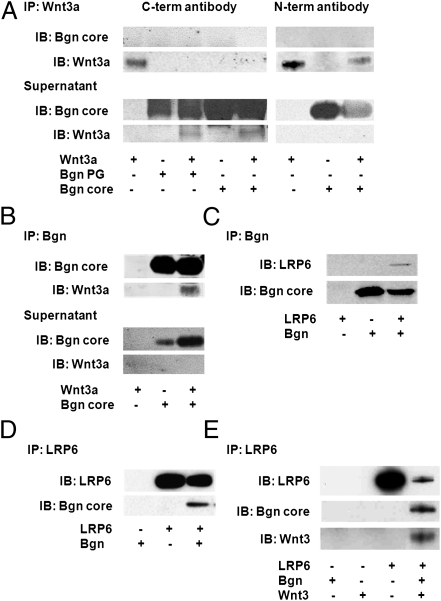
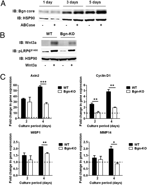
Although extracellular control of canonical Wnt signaling is crucial for tissue homeostasis, the role of the extracellular microenvironment in modulating this signaling pathway is largely unknown. In the present study, we show that a member of the small leucine-rich proteoglycan family, biglycan, enhances canonical Wnt signaling by mediating Wnt function via its core protein. Immunoprecipitation analysis revealed that biglycan interacts with both the canonical Wnt ligand Wnt3a and the Wnt coreceptor low-density lipoprotein receptor-related protein 6 (LRP6), possibly via the formation of a trimeric complex. Biglycan-deficient cells treated with exogenous Wnt3a had less Wnt3a retained in cell layers compared with WT cells. Furthermore, the Wnt-induced levels of LRP6 phosphorylation and expression of several Wnt target genes were blunted in biglycan-deficient cells. Both recombinant biglycan proteoglycan and biglycan core protein increased Wnt-induced β-catenin/T cell-specific factor-mediated transcriptional activity, and this activity was completely inhibited by Dickkopf 1. Interestingly, recombinant biglycan was able to rescue impaired Wnt signaling caused by a previously described missense mutation in the extracellular domain of human LRP6 (R611C). Furthermore, biglycan's modulation of canonical Wnt signaling affected the functional activities of osteoprogenitor cells, including the RUNX2-mediated transcriptional activity and calcium deposition. Use of a transplant system and a fracture healing model revealed that expression of Wnt-induced secreted protein 1 was decreased in bone formed by biglycan-deficient cells, further suggesting reduced Wnt signaling in vivo. We propose that biglycan may serve as a reservoir for Wnt in the pericellular space and modulate Wnt availability for activation of the canonical Wnt pathway.


Conflict of interest statement

The authors declare no conflict of interest.

Figures

Fig. 1.
Fig. 1.
Biglycan interacts with Wnt3a and LRP6 via its core protein. (A and B) Immunoblotting with biglycan or Wnt3a antibodies of immunoprecipitates (IPs) collected using biglycan and Wnt3a antibodies after mixing of recombinant human biglycan (with and without glycanation) with recombinant human Wnt3a protein. (A) IP using antibodies to the C terminus of Wnt3a (Left) or with antibodies to the N-terminal region of Wnt3a (Right). (B) IP using antibodies to Bgn. (C and D) IPs of cell lysates of HEK-293T cells overexpressing V5-tagged human biglycan and V5-tagged human LRP6 using antibodies for biglycan (C) or LRP6 (D), followed by immunoblotting of pull-down lysates with V5-specific antibodies. (E) IPs of cell lysates of HEK-293T cells overexpressing V5-tagged human biglycan, V5-tagged human LRP6, and HA-tagged human/mouse Wnt3 using antibodies for LRP6, followed by immunoblotting of pull-down lysates with V5- and HA-specific antibodies.
Fig. 2.
Fig. 2.
Biglycan-deficient calvarial cells retain less Wnt3a in cell layers and demonstrate both reduced Wnt-induced phosphorylation of LRP6 and reduced β-catenin/TCF-mediated transcriptional activity. (A) Immunoblot analysis for biglycan core protein after treatment of an equal volume of lysate proteins obtained from WT calvarial cell layers with chondroitinase ABC. (B) Immunoblot analysis for Wnt3a and phosphorylation of LRP6 (pLRP6S1490) on protein extracts obtained from WT and biglycan-deficient calvarial cells after treatment with Wnt3a. (C) Relative expression levels of Wnt target genes measured by real-time RT-PCR in WT and biglycan-deficient calvarial cells after treatment with Wnt3a. Values represent mean relative expression ± SD (n = 3), shown as Wnt3a-induced fold change in gene expression. Gene expression was normalized to untreated controls at each time point (indicated by the dotted line). *P < 0.05; **P < 0.01; ***P < 0.001.
Fig. 3.
Fig. 3.
Both biglycan proteoglycan and biglycan core protein enhance Wnt-induced β-catenin/TCF-mediated transcriptional activity via LRP6, and this activity is inhibited by Dkk1. (A) Effect of biglycan proteoglycan (PG) and biglycan core protein on Wnt3a-induced reporter activity in β-catenin reporter cells overexpressing LRP6-WT or inactive LRP6-MT. Values represent mean relative luminescence ± SD (n = 3). *P < 0.05; **P < 0.01; ***P < 0.001, control (no biglycan) vs. biglycan treatment. (B) Effect of biglycan PG and biglycan core protein on Wnt3a-induced reporter activity in β-catenin reporter cells overexpressing LRP6-MT. Values represent mean relative luminescence ± SD (n = 3). *P < 0.05; **P < 0.01, control (no biglycan) vs. biglycan treatment. (C) Effect of Dkk1 on Wnt3a-induced reporter activity in β-catenin reporter cells in the presence of biglycan core protein. Values represent mean relative luminescence ± SD (n = 3). *P < 0.05, +Wnt3a/−biglycan core vs. +Wnt3a/+biglycan core at 0 and 10 ng/mL Dkk1. (D) Effect of LiCl-induced β-catenin reporter activity in β-catenin reporter cells in the presence of biglycan core protein. NaCl was used for control purposes. Values represent mean relative luminescence ± SD (n = 3).
Fig. 4.
Fig. 4.
Biglycan core protein rescues impaired β-catenin/TCF-mediated transcriptional activity caused by the R611C missense mutation in LRP6 and stimulates Wnt-induced RUNX2 transcriptional activity. (A) Location of the R611C mutation in LRP6. (B) Effect of biglycan core protein on Wnt3a-induced β-catenin reporter activity in β-catenin reporter cells overexpressing LRP6-WT or LRP6-R611C. Values represent the mean relative luminescence ± SD (n = 3). *P < 0.05; **P < 0.01, LRP6-R611C (no biglycan) vs. LRP6-R611C (biglycan-treated) and LRP6-WT (no biglycan). (C) Wnt3a-induced β-catenin reporter activity in cells expressing only endogenous LRP6 (indicated by LRP6-MT) compared with cells overexpressing LRP6-WT or LRP6-R611C. Values represent mean relative luminescence ± SD (n = 3). *P < 0.05; **P < 0.01. (D and E) Immunodetection of the V5 tag (fused with biglycan, LRP6-WT, and LRP6-R611C) of IPs collected using biglycan (D) or LRP6 (E) antibodies after harvesting of β-catenin reporter cells overexpressing biglycan, LRP6-WT, LRP6-R611C, or a combination of these. (F) Effect of biglycan core protein on Wnt3a-induced p6OSE2-luc reporter activity in MC3T3-E1 cells. Values represent mean relative luminescence ± SD (n = 6). **P < 0.01.
Fig. 5.
Fig. 5.
Biglycan-deficient cells have reduced Wnt-induced mineralization in cultures and show less WISP1 expression in trabecular structures formed in transplants and during fracture healing. (A) Wnt-induced calcium deposition assessed by alizarin red staining of WT and biglycan-deficient BMSCs. Values represent mean relative alizarin red staining ± SD (n = 3). *P < 0.05; **P < 0.01. (B) Micro-CT images of WT (Upper) and biglycan-deficient (Lower) transplants and quantitative assessment of the bone mineral density of trabecular structures. (C) H&E staining of sections of WT (Upper) and biglycan-deficient (Lower) transplants. (D) Immunostaining for WISP1 on sections through the center of WT (Upper) and biglycan-deficient (Lower) transplants. Arrows indicate positive staining in the trabecular-like bone formed in the transplants. (E) H&E staining of sections of WT (Upper) and biglycan-deficient (Lower) femurs at 14 d postfracture. White arrows indicate the fracture area. (F) Immunostaining for WISP1 on sections through WT (Upper) and biglycan-deficient (Lower) femurs at 14 d postfracture showing WISP1 expression surrounding the woven bone formed in the callus area. (G) Relative WISP1 gene expression levels measured by real-time RT-PCR in WT and biglycan-deficient callus areas of femurs at 7 d postfracture. Values represent mean relative expression ± SD (n = 4). *P < 0.05.

References

    1. MacDonald BT, Tamai K, He X. Wnt/β-catenin signaling: Components, mechanisms, and diseases. Dev Cell. 2009;17:9–26. - PMC - PubMed
    1. Bienz M, Clevers H. Linking colorectal cancer to Wnt signaling. Cell. 2000;103:311–320. - PubMed
    1. Patel MS, Karsenty G. Regulation of bone formation and vision by LRP5. N Engl J Med. 2002;346:1572–1574. - PubMed
    1. Lin X. Functions of heparan sulfate proteoglycans in cell signaling during development. Development. 2004;131:6009–6021. - PubMed
    1. Ai X, et al. QSulf1 remodels the 6-O sulfation states of cell surface heparan sulfate proteoglycans to promote Wnt signaling. J Cell Biol. 2003;162:341–351. - PMC - PubMed

Publication types

MeSH terms

LinkOut - more resources