. 2011 Oct 18;108(42):17444-9.
doi: 10.1073/pnas.1107023108. Epub 2011 Oct 3.

Macrophage migration inhibitory factor (MIF) exerts antifibrotic effects in experimental liver fibrosis via CD74

Affiliations

Macrophage migration inhibitory factor (MIF) exerts antifibrotic effects in experimental liver fibrosis via CD74

Daniel Heinrichs et al. Proc Natl Acad Sci U S A. .

Abstract

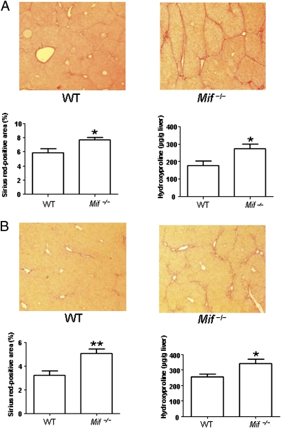
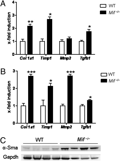
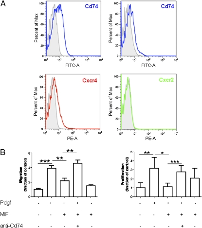
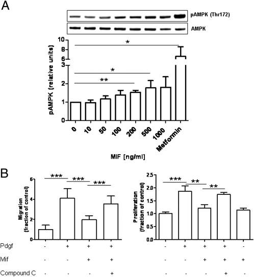
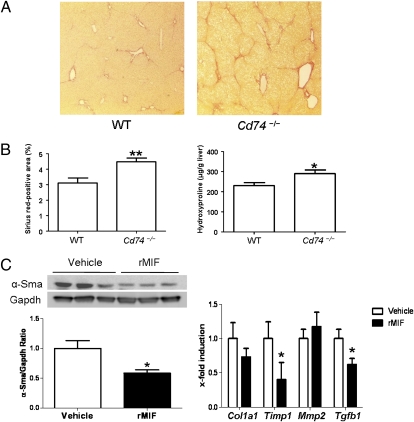
Macrophage migration inhibitory factor (MIF) is a pleiotropic inflammatory cytokine that has been implicated in various inflammatory diseases. Chronic inflammation is a mainstay of liver fibrosis, a leading cause of morbidity worldwide, but the role of MIF in liver scarring has not yet been elucidated. Here we have uncovered an unexpected antifibrotic role for MIF. Mice genetically deleted in Mif (Mif(-/-)) showed strongly increased fibrosis in two models of chronic liver injury. Pronounced liver fibrosis in Mif(-/-) mice was associated with alterations in fibrosis-relevant genes, but not by a changed intrahepatic immune cell infiltration. Next, a direct impact of MIF on hepatic stellate cells (HSC) was assessed in vitro. Although MIF alone had only marginal effects on HSCs, it markedly inhibited PDGF-induced migration and proliferation of these cells. The inhibitory effects of MIF were mediated by CD74, which we detected as the most abundant known MIF receptor on HSCs. MIF promoted the phosphorylation of AMP-activated protein kinase (AMPK) in a CD74-dependent manner and, in turn, inhibition of AMPK reversed the inhibition of PDGF-induced HSC activation by MIF. The pivotal role of CD74 in MIF-mediated antifibrotic properties was further supported by augmented liver scarring of Cd74(-/-) mice. Moreover, mice treated with recombinant MIF displayed a reduced fibrogenic response in vivo. In conclusion, we describe a previously unexplored antifibrotic function of MIF that is mediated by the CD74/AMPK signaling pathway in HSCs. The results imply MIF and CD74 as targets for treatment of liver diseases.


Conflict of interest statement

The authors declare no conflict of interest.

Figures

Fig. 1.
Fig. 1.
Enhanced liver fibrosis in Mif−/− mice in two independent experimental models. (A) CCl4 model: representative Sirius red stainings (Upper) of WT and Mif−/− mice after challenge with CCl4 for 6 wk. Sirius red stainings from 12 mice per group were quantitated (Lower Left). Increased fibrosis in Mif−/− mice compared with WT (n = 12 per group) was further validated by significantly increased concentrations of hydroxyproline (Lower Right). (B) TAA model: challenge with TAA for 6 wk of WT vs. Mif−/− mice. Exaggerated fibrosis in Mif−/− mice compared with WT (n = 12 per group) is evident by representative Sirius red staining (Upper) and quantification of Sirius red-positive area (Lower Left) and increased concentrations of hydroxyproline (Lower Right). Asterisks indicate statistical significance: *P < 0.05, **P < 0.01. (Magnification: A and B, 200×.)
Fig. 2.
Fig. 2.
Increased expression of HSC-related genes and α-Sma in Mif−/− mice. (A) CCl4 treatment of Mif−/− mice leads to significantly increased mRNA expression of Col1a1, Timp1, and Tgfb1 compared with WT mice. (B) Same as in A, except that fibrosis was induced by TAA treatment. Asterisks indicate statistical significance: *P < 0.05, **P < 0.01, ***P < 0.001. (C) Augmented HSC activation after CCl4 challenge in Mif−/− mice (lanes 5–8) compared with WT (lanes 1–4) is further evidenced by increased expression of α-Sma protein expression (Western blot from total liver lysates; loading control: Gapdh). Each lane represents a cell lysate from an independent mouse.
Fig. 3.
Fig. 3.
Surface expression and role of CD74 in regulation of HSCs by MIF. (A) Expression of MIF receptors on immortalized and primary HSCs. FACS analysis reveals marked expression of CD74 (Upper Left: immortalized murine HSCs, Upper Right: primary murine HSCs) and low-to-moderate expression CXCR4 on stellate cells. CXCR2 is not expressed on HSCs. (B) Chemotaxis (Left) and BrdU incorporation-based proliferation experiments (Right) experiments reveal that MIF inhibits PDGF-induced HSC behavior, but MIF alone does not have substantial effects on these cells. Note, that the inhibitory effects of MIF on PDGF-induced HSC activation can be completely blocked by pretreatment of the cells with neutralizing CD74 antibody (anti-CD74). Each type of incubation was performed at least twice in quadruplicate each. Asterisks indicate statistical significance: *P < 0.05, **P < 0.01, ***P < 0.001.
Fig. 4.
Fig. 4.
Regulation of hepatic stellate cell activity by MIF is mediated by AMPK activation. (A) Recombinant MIF induces a dose-dependent phosphorylation of AMPK at position Thr-172. A representative Western blot is shown. The bar diagram represents mean values of three independent experiments. (B) Inhibition of PDGF-induced HSC migration and proliferation by MIF is mediated by AMPK activation. HSCs were incubated with PDGF and MIF in the presence or absence of the AMPK inhibitor Compound C, which significantly reverts the inhibitory effects of MIF. All types of incubation were performed at least twice in quadruplicate each. Asterisks indicate statistical significance: *P < 0.05, ***P < 0.001.
Fig. 5.
Fig. 5.
Enhanced liver fibrosis in Cd74−/− mice in CCl4 fibrosis model. (A) Representative Sirius red stainings of WT and Cd74−/− mice after challenge with CCl4. (Magnification: 200×.) (B) Increased fibrosis in Cd74−/− mice (n = 8 per group) as validated by quantification of Sirius red-positive areas (Left) and hydroxyproline content (Right). (C) Daily administration of recombinant MIF represses the CCl4 induced activation of stellate cells as assessed by total α-Sma protein expression. Three representative samples of each group and the overall ratio of α-Sma/Gapdh are displayed. (D) The reduced fibrogenic response in mice treated with rMIF is validated by repression of fibrosis related-genes compared with vehicle-treated mice. *P < 0.05, **P < 0.01.

Comment in

References

    1. Schuppan D, Afdhal NH. Liver cirrhosis. Lancet. 2008;371:838–851. - PMC - PubMed
    1. El-Serag HB, Mason AC. Rising incidence of hepatocellular carcinoma in the United States N Engl. J Med. 1999;340:745–750. - PubMed
    1. Ginès P, Cárdenas A, Arroyo V, Rodés J. Management of cirrhosis and ascites. N Engl J Med. 2004;350:1646–1654. - PubMed
    1. Friedman SL. Mechanisms of hepatic fibrogenesis. Gastroenterology. 2008;134:1655–1669. - PMC - PubMed
    1. Friedman SL. Hepatic stellate cells: Protean, multifunctional, and enigmatic cells of the liver. Physiol Rev. 2008;88(1):125–172. - PMC - PubMed

Publication types

MeSH terms

Substances

LinkOut - more resources