. 2011 Oct 18;108(42):17438-43.
doi: 10.1073/pnas.1111855108. Epub 2011 Oct 3.

Animal-specific C-terminal domain links myeloblastosis oncoprotein (Myb) to an ancient repressor complex

Affiliations

Animal-specific C-terminal domain links myeloblastosis oncoprotein (Myb) to an ancient repressor complex

Laura Andrejka et al. Proc Natl Acad Sci U S A. .

Abstract

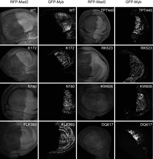
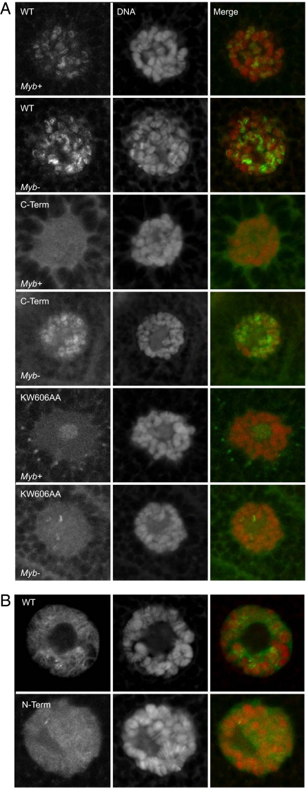
Members of the Myb oncoprotein and E2F-Rb tumor suppressor protein families are present within the same highly conserved multiprotein transcriptional repressor complex, named either as Myb and synthetic multivuval class B (Myb-MuvB) or as Drosophila Rb E2F and Myb-interacting proteins (dREAM). We now report that the animal-specific C terminus of Drosophila Myb but not the more highly conserved N-terminal DNA-binding domain is necessary and sufficient for (i) adult viability, (ii) proper localization to chromosomes in vivo, (iii) regulation of gene expression in vivo, and (iv) interaction with the highly conserved core of the MuvB/dREAM transcriptional repressor complex. In addition, we have identified a conserved peptide motif that is required for this interaction. Our results imply that an ancient function of Myb in regulating G2/M genes in both plants and animals appears to have been transferred from the DNA-binding domain to the animal-specific C-terminal domain. Increased expression of B-MYB/MYBL2, the human ortholog of Drosophila Myb, correlates with poor prognosis in human patients with breast cancer. Therefore, our results imply that the specific interaction of the C terminus of Myb with the MuvB/dREAM core complex may provide an attractive target for the development of cancer therapeutics.


Conflict of interest statement

The authors declare no conflict of interest.

Figures

Fig. 1.
Fig. 1.
Mutants of Drosophila Myb. (A) (Top) Cartoon of the WT protein. The cyan boxes indicate the three conserved Myb repeats that constitute the paneukaryotic N-terminal DNA-binding domain. The red box indicates the conserved animal-specific C-terminal domain. The lines below the cartoon indicate the location of 17 previously undescribed alanine substitution mutants. Also shown are cartoons of the N-terminal (Middle; N-term) mutant (residues 1–316) and C-terminal (Bottom; C-term) mutant (residues 247–657). (B) Alignment of a conserved animal-specific C-terminal region that includes a previously described heat-sensitive hypomorphic Myb1 mutant was made using MACAW software (NCBI) (34, 51). The three Myb genes of vertebrates arose via duplications that occurred before the divergence of all species examined thus far and are represented here by human MYB/C-MYB, MYBL1/A-MYB, and MYBL2/B-MYB (11).
Fig. 2.
Fig. 2.
Rescue of Myb-null Drosophila by Myb WT and mutant proteins. (Upper) Rescue of adult viability of the otherwise lethal MybMH107-null mutant via the GAL4-UAS binary system at 25 °C. (Lower) Rescue of adult viability of the otherwise lethal MybMH107-null mutant via the native Myb promoter. Details of the Drosophila genetics are provided in Figs. S1 and S4. ND, indicated mutant not tested for rescue at the indicated temperature.
Fig. 3.
Fig. 3.
Regulation of Mad2 expression by Myb WT and mutant proteins. The indicated Myb proteins were expressed via engrailed-GAL4 in the posterior compartments of third-instar MybMH107-null mutant larvae. Imaginal disks were dissected, fixed, stained with anti-RFP antibodies, and visualized by laser scanning confocal microscopy at 400× magnification. Each row displays RFP-Mad2 (columns 1 and 3) and GFP-Myb (columns 2 and 4) in two different imaginal wing disks.
Fig. 4.
Fig. 4.
Chromosomal localization of Myb WT and mutant proteins. (A) Indicated GFP-Myb fusion proteins were expressed via the native Myb promoter in either Myb WT (Myb+) or Myb-null (Myb) larvae. Salivary glands were dissected, fixed, and visualized by laser scanning confocal microscopy at 600× magnification. Each row displays GFP-Myb (Left), DNA staining (Center), and merged images (Right) of the same salivary gland nucleus. The central area of lightly stained DNA visible in some nuclei is the nucleolus. (B) Full-length WT or N-terminal GFP-Myb protein was expressed via daughterless-GAL4.
Fig. 5.
Fig. 5.
Coprecipitation of the MuvB core complex by Myb WT and mutant proteins. FLAG-tagged Myb WT or mutant protein was expressed in Drosophila S2 cells by transient DNA transfection. RNAi directed against the 5′-UTR was used to knock down endogenous Myb expression. Immunoblots of total extracts (input) or of immunoprecipitated complexes (FLAG-IP) were analyzed using the indicated antibodies. (Left) Analysis of interaction with the MuvB core complex by full-length (WT), N-terminal, or C-terminal Myb. The anti-Myb antibody recognizes the N-terminal repeats of the Myb DNA-binding domain; therefore, it cannot detect the C-terminal mutant (19). A 50-kDa background band detected by anti-FLAG in all FLAG-IP samples is mouse Ig-heavy chain that is released from the anti-FLAG beads used for immunoprecipitation. (Right) Analysis of interaction with the MuvB core complex by WT or the indicated alanine substitution mutants of Myb.
Fig. 6.
Fig. 6.
Function and evolutionary conservation of Myb and MuvB core proteins. (Left) Model for the function of WT and mutant Myb proteins in gene regulation. The MuvB core and associated proteins repress gene expression. This repression is relieved by contact with the animal-specific C terminus of Myb. The N-terminal mutant of Myb acts as a dominant loss-of-function allele, presumably by blocking access by the WT Myb protein. (Right) Evolutionary conservation of Myb and MuvB core proteins. MYB-N indicates a Myb DNA-binding domain closely related to that of animal Myb proteins. MYB-C indicates the animal-specific C-terminal domain. The core MuvB components are indicated by their names in Drosophila and in C. elegans. Homologous protein sequences were identified by searching the NCBI databases using TBlastn. Black squares indicate the presence of a homologous protein. Light gray squares indicate the absence of a homologous protein in the current NCBI database.

References

    1. Lipsick JS, Wang DM. Transformation by v-Myb. Oncogene. 1999;18:3047–3055. - PubMed
    1. Ramsay RG, Gonda TJ. MYB function in normal and cancer cells. Nat Rev Cancer. 2008;8:523–534. - PubMed
    1. Klempnauer KH, Symonds G, Evan GI, Bishop JM. Subcellular localization of proteins encoded by oncogenes of avian myeloblastosis virus and avian leukemia virus E26 and by chicken c-myb gene. Cell. 1984;37:537–547. - PubMed
    1. Boyle WJ, Lampert MA, Lipsick JS, Baluda MA. Avian myeloblastosis virus and E26 virus oncogene products are nuclear proteins. Proc Natl Acad Sci USA. 1984;81:4265–4269. - PMC - PubMed
    1. Biedenkapp H, Borgmeyer U, Sippel AE, Klempnauer KH. Viral myb oncogene encodes a sequence-specific DNA-binding activity. Nature. 1988;335:835–837. - PubMed

Publication types

MeSH terms

LinkOut - more resources