. 2012 Jan;11(1):M111.012054.
doi: 10.1074/mcp.M111.012054. Epub 2011 Oct 3.

Evolutionary diversification of Mesobuthus α-scorpion toxins affecting sodium channels

Affiliations

Evolutionary diversification of Mesobuthus α-scorpion toxins affecting sodium channels

Shunyi Zhu et al. Mol Cell Proteomics. 2012 Jan.

Abstract

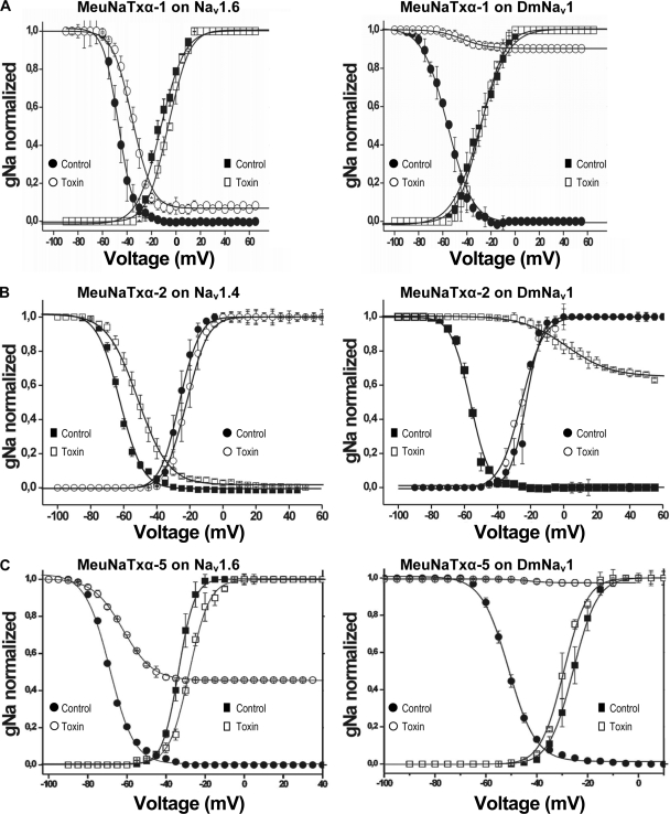
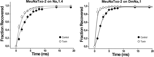
α-Scorpion toxins constitute a family of peptide modulators that induce a prolongation of the action potential of excitable cells by inhibiting voltage-gated sodium channel inactivation. Although they all adopt a conserved structural scaffold, the potency and phylogentic preference of these toxins largely vary, which render them an intriguing model for studying evolutionary diversification among family members. Here, we report molecular characterization of a new multigene family of α-toxins comprising 13 members (named MeuNaTxα-1 to MeuNaTxα-13) from the scorpion Mesobuthus eupeus. Of them, five native toxins (MeuNaTxα-1 to -5) were purified to homogeneity from the venom and the solution structure of MeuNaTxα-5 was solved by nuclear magnetic resonance. A systematic functional evaluation of MeuNaTxα-1, -2, -4, and -5 was conducted by two-electrode voltage-clamp recordings on seven cloned mammalian voltage-gated sodium channels (Na(v)1.2 to Na(v)1.8) and the insect counterpart DmNa(v)1 expressed in Xenopus oocytes. Results show that all these four peptides slow inactivation of DmNa(v)1 and are inactive on Na(v)1.8 at micromolar concentrations. However, they exhibit differential specificity for the other six channel isoforms (Na(v)1.2 to Na(v)1.7), in which MeuNaTxα-4 shows no activity on these isoforms and thus represents the first Mesobuthus-derived insect-selective α-toxin identified so far with a half maximal effective concentration of 130 ± 2 nm on DmNa(v)1 and a half maximal lethal dose of about 200 pmol g(-1) on the insect Musca domestica; MeuNaTxα-2 only affects Na(v)1.4; MeuNaTxα-1 and MeuNaTxα-5 have a wider range of channel spectrum, the former active on Na(v)1.2, Na(v)1.3, Na(v)1.6, and Na(v)1.7, whereas the latter acting on Na(v)1.3-Na(v)1.7. Remarkably, MeuNaTxα-4 and MeuNaTxα-5 are two nearly identical peptides differing by only one point mutation at site 50 (A50V) but exhibit rather different channel subtype selectivity, highlighting a switch role of this site in altering the target specificity. By the maximum likelihood models of codon substitution, we detected nine positively selected sites (PSSs) that could be involved in functional diversification of Mesobuthus α-toxins. The PSSs include site 50 and other seven sites located in functional surfaces of α-toxins. This work represents the first thorough investigation of evolutionary diversification of α-toxins derived from a specific scorpion lineage from the perspectives of sequence, structure, function, and evolution.


Conflict of interest statement

The authors have declared no conflicts of interest.

Figures

Fig. 1.
Fig. 1.
The multigene family of α-toxins in M. eupeus. A, Strategy for isolating cDNAs of a complete α-toxin multigene family in M. eupeus by combination of different primers (represented by arrows) whose sequence information and pairs for PCR amplification are provided in supplemental Table S1 and S2. Different regions of the tailed first-strand cDNA encoding an α-toxin are shown in color. UTR, untranslational region. SP, signal peptide. MP, mature peptide; B, Multiple sequence alignment of α-toxins. Cysteines are shadowed in yellow and residues conserved across the alignment in color (Basic: blue; hydrophobic: green; polar: cyan). Extra C-terminal amino acids are underlined once and amidated residues are boldfaced. Secondary structure elements (arrow: β-strand; cylinder: α-helix) and four disulfide bridges (indicated by lines) extracted from the NMR structure of MeuNaTxα-5 are shown at the bottom of the alignment. PSSs predicted from the codon-substitution model are indicated by red arrows. N-turn and four loops (J, M, B, and F) are shown at the top of the alignment. g, m, and p represent genomic, mRNA and protein, respectively. * indicates MeuNaTxα, where native peptides were isolated from the venom.
Fig. 2.
Fig. 2.
Exon-intron structure of M. eupeus α-toxins. A, Genomic organization conservation among MeuNaTxα-1, MeuNaTxα-2 and MeuNaTxα-5′. Signal peptide sequences are italicized and residues split by a phase I intron are boxed. Identical amino acids and nucleotides are shadowed in gray. Extra residues are underlined once; B, Conservation of 5′- and 3′-splicing sites of introns.
Fig. 3.
Fig. 3.
Phylogenetic tree of Mesobuthus α-toxin genes. The tree is a bootstrap consensus tree based upon 1000 replicates of the Neighbor-Joining (NJ) algorithm with the Maximum Composite Likelihood model of DNA substitutions. The scale bar shows total nucleotide divergence. > 50% bootstrap values are shown at nodes. Branches derived from M. eupeus are in red. Similar tree topology was obtained with the Minimum Evolution (ME) or Unweighted Pair Group Method Algorithm (UPGMA) (data not shown).
Fig. 4.
Fig. 4.
Purification of scorpion α-toxins from the M. eupeus crude venom by RP-HPLC. Fractions corresponding to MeuNaTxα-1 - MeuNaTxα-5 and those to three known peptides (MeuTXKα1, MeuTx3B and BeKm-1) (23) are indicated by arrows. The Agilent semi-prep Zorbax 300SB-C18 (9.2 × 250 mm, 5 μm) was equilibrated with 0.1% TFA in water (v/v) and peptide components were eluted from the column with a linear gradient from 0 to 60% acetonitrile in 0.1% TFA in water (v/v) within 60 min with a flow rate of 1 ml/min. The UV absorbance trace was followed at 225 nm.
Fig. 5.
Fig. 5.
Solution structure of MeuNaTxα-5. A, Stereo view of the 20 NMR models, superimposed for a minimum r.m.s.d. to the (C, Cα, N) atoms from residues Ala1 to His66; B, Ribbon representation of the toxin. Disulfide bridges (SS1-SS4) are shown in a blue stick model. Secondary structure regions are assigned by STRIDE (http://webclu.bio.wzw.tum.de/stride/). Beginning and ending amino acids of each secondary structure element (α-helix in red and β-strand in cyan) are labeled according to the peptide sequence.
Fig. 6.
Fig. 6.
Differential effects of MeuNaTxα-1, -2, -4, and -5 on Nav isoforms expressed in X. leavis oocytes. Representative whole cell Na+ current traces of oocytes expressing cloned Nav isoforms (Nav1.2–Nav1.8, and DmNav1) are shown. The dotted line indicates the zero-current level. The * indicates the steady-state current peak amplitude in the presence of 1–2 μm toxin. CNS: central nervous system; PNS: peripheral nervous system.
Fig. 7.
Fig. 7.
Dose-response curves of M. eupeus α-toxins. The curves were obtained by plotting the relative I30 ms/Ipeak values of the channels in function of the toxin concentrations. All the curves were fitted with the Hill equation. A, MeuNaTxα-1 on DmNav1 and Nav1.6; B, MeuNaTxα-2 on DmNav1 and Nav1.4; C, MeuNaTxα-4 on DmNav1; D, MeuNaTxα-5 on DmNav1 and Nav1.6.
Fig. 8.
Fig. 8.
Effects of M. eupeus α-toxins on the voltage dependence of activation and steady-state inactivation curves of Nav channels. The steady-state currents of the channels in control and after the addition of 1 μm toxin are shown. A, MeuNaTxα-1: Nav1.6 (left) and DmNav1 (right); B, MeuNaTxα-2: Nav1.4 (left) and DmNav1 (right); C, MeuNaTxα-5: Nav1.6 (left) and DmNav1 (right).
Fig. 9.
Fig. 9.
Effects of MeuNaTxα-2 on the recovery from fast inactivation of Nav channels. Nav1.4 (left) and DmNav1 (right) in control and in the presence of 1 μm peptide are shown.
Fig. 10.
Fig. 10.
Pairwise ω estimation of Mesobuthus α-toxins. The ω values, estimated by the method of Nei and Gojobori (28) implemented in CODEML (29), are shadowed in different colors: ω<0.5 (gray); 0.5≤ω≤1 (yellow); 1 < ω < 10 (red); ∞ (blue).
Fig. 11.
Fig. 11.
Structural and functional features of PSSs in Mesobuthus α-toxins. A, Mapping of the PSSs on the crystal structure of BmK M1 (pdb entry 1SN1), where PSSs are in shown in red. Different amino acid types in PSSs are highlighted in color: blue, positively charged; red, negatively charged; cyan, polar; green, hydrophobic; B, Comparison of functional sites (FSs) of four well-identified α-toxins and PSSs estimated from different sequence sources [2004 (52); 2010 (47)]. βT: β-turn; NT: N-terminal five-residue turn; “o” represents positions of FSs or PSSs. PSSs commonly predicted in three different studies are shadowed in yellow and PSSs reported here associated with functions are shown in red. *residue numberings in Lqh3 are 39, 45, 59, 64, and 66, respectively. Identical FSs or PSSs are boxed.
Fig. 12.
Fig. 12.
Molecular basis for channel subtype selectivity of MeuNaTxαs. A, The model structure of DI-DIV domains of DmNav1. All the four domains, shown in different colors (DI, green; DII, purple; DIII, blue; DIV, red), were build by Homer, a comparative modeling server (http://protein.cribi.unipd.it/homer/) and the atomic structure of a mammalian chimeric VGPC (pdb entry 2R9R) was used as template. Domain assembly was performed by Swiss PDB Viewer (http://spdbv.vital-it.ch/); B, Structural representation of the DmNav1 site 3 comprising three loops connecting S3 and S4 in domain IV (LDIV:S3-S4) shown as molecular surface, and S5-S6 in DI and DIV (LDI:S5-S6, LDIV:S5-S6); C, Toxin sensitivity of different VGSCs associated with sequences in LDIV:S3-S4. α: MeuNaTxα. Identical amino acids to DmNav1 are shadowed in yellow. The phylogeny was reconstructed based on the sequence of site 3 listed here. Bootstrap values are shown at nodes. +, activity; -, no activity.

Similar articles

Cited by

References

    1. Rochat H., Martin-Eauclaire M. F. (2000) Animal Toxins: Facts and Protocols. Birkhauser Verlag, Basel, Boston, Berlin
    1. Billen B., Bosmans F., Tytgat J. (2008) Animal peptides targeting voltage-activated sodium channels. Curr. Pharm. Design. 14, 2492–2502 - PubMed
    1. Lewis R. J., Garcia M. L. (2003) Therapeutic potential of venom peptides. Nat. Rev. Drug Discov. 2, 790–802 - PubMed
    1. Catterall W. A. (1995) Structure and function of voltage-gated ion channels. Annu. Rev. Biochem. 64, 493–531 - PubMed
    1. Catterall W. A. (2000) From ionic currents to molecular mechanisms: The structure and function of voltage-gated sodium channels. Neuron 26, 13–25 - PubMed

Publication types

LinkOut - more resources