. 2011;6(9):e23059.
doi: 10.1371/journal.pone.0023059. Epub 2011 Sep 28.

Spinal astrocytic activation is involved in a virally-induced rat model of neuropathic pain

Affiliations

Spinal astrocytic activation is involved in a virally-induced rat model of neuropathic pain

Gui-He Zhang et al. PLoS One. 2011.

Abstract

Postherpetic neuralgia (PHN), the most common complication of herpes zoster (HZ), plays a major role in decreased life quality of HZ patients. However, the neural mechanisms underlying PHN remain unclear. Here, using a PHN rat model at 2 weeks after varicella zoster virus infection, we found that spinal astrocytes were dramatically activated. The mechanical allodynia and spinal central sensitization were significantly attenuated by intrathecally injected L-α-aminoadipate (astrocytic specific inhibitor) whereas minocycline (microglial specific inhibitor) had no effect, which indicated that spinal astrocyte but not microglia contributed to the chronic pain in PHN rat. Further study was taken to investigate the molecular mechanism of astrocyte-incudced allodynia in PHN rat at post-infection 2 weeks. Results showed that nitric oxide (NO) produced by inducible nitric oxide synthase mediated the development of spinal astrocytic activation, and activated astrocytes dramatically increased interleukin-1β expression which induced N-methyl-D-aspartic acid receptor (NMDAR) phosphorylation in spinal dorsal horn neurons to strengthen pain transmission. Taken together, these results suggest that spinal activated astrocytes may be one of the most important factors in the pathophysiology of PHN and "NO-Astrocyte-Cytokine-NMDAR-Neuron" pathway may be the detailed neural mechanisms underlying PHN. Thus, inhibiting spinal astrocytic activation may represent a novel therapeutic strategy for clinical management of PHN.


Conflict of interest statement

Competing Interests: The authors have declared that no competing interests exist.

Figures

Figure 1
Figure 1. Experimental procedures in this study (A) and mechanical allodynia in VZV infected rats (B and C).
(A) The timeline represents the period during which behavior, histochemistry, PCR and Western blot studies were performed per week after VZV infection. Intrathecal catheterization was performed on rats and followed by 3-day recovery. The pharmacology, electrophysiology, and NOS studies were conducted at post-infection 2 weeks when the mechanical allodynia reached the highest level. (B) Compared with Naive rats and Mock infected rats, the paw withdrawal threshold of VZV infected rats was significantly decreased. * P<0.05, ** P<0.01 vs. Naive rats and Mock infected rats. (C) Systemic treatment with antiviral agent valaciclovir had no effect on the development of mechanical allodynia. * P<0.05, ** P<0.01 vs. Mock infected rats. All data were calculated as mean ± SEM (n = 10/group/week).
Figure 2
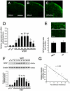
Figure 2. Spinal astrocytes were activated in VZV infected rats, which was significantly correlated to mechanical allodynia.
(A–C) Compared with Naive rats and Mock infected rats, GFAP-like immunoreactivity (-LI) in spinal dorsal horn of VZV infected rats was significantly increased. Bar = 200 µm. (D) Real-time RT-PCR showed that spinal mRNA expression of GFAP was significantly upregulated in VZV infected rats compared to naive rats and mock infected rats. (E) With regard to the total number of GFAP positive cells, there was no difference between Naïve rats and VZV infected rats. Bar = 10 µm. (F) Compared to Naive rats and Mock infected rats, Western blot analysis showed that spinal GFAP expression was significantly increased in VZV infected rats. (G) The expression level of GFAP was found to be significantly correlated to the paw withdrawal threshold in VZV infected rats (P<0.001, r = −0.868). All data were calculated as mean ± SEM (n = 10/group/week). * P<0.05, ** P<0.01 vs. Naive rats and Mock infected rats; ΔP<0.05 vs. post-infection 2 weeks rats.
Figure 3
Figure 3. Spinal microglia was not activated in VZV infected rats, astrocytic specific inhibitor LAA but not microglial specific inhibitor minocycline could attenuate mechanical allodynia.
(A) No significant difference in OX42 mRNA expression in spinal cord was observed among naive rats, mock infected rats and VZV infected rats. (B) With regard to OX42-like immunoreactivity (-LI) in spinal dorsal horn, there was no difference between Naïve rats and VZV infected rats. Bar = 200 µm. (C) With regard to OX42 expression in spinal cord, there was no difference among Naive rats, Mock infected rats and VZV infected rats. In VZV infected rats, OX42 expression was unchanged through the period tested. (D) Intrathecal injection of LAA significantly attenuated the allodynia. However, minocycline did not influence the allodynia. All data were calculated as mean ± SEM (n = 10/group). * P<0.05, ** P<0.01 vs. VZV+Saline group or VZV+minocycline group in D.
Figure 4
Figure 4. Astrocytic activation contributed to spinal central sensitization in VZV infected rats.
Comparative recordings of responsiveness of spinal dorsal horn wide dynamic range (WDR) neurons to mechanical (brush, pressure and pinch) stimuli in Mock infected group (A), VZV infected group (B), VZV+LAA group (C) and VZV+minocycline group (D). (A–D) The responsiveness of WDR neurons was gradedly increased with the increase in mechanical intensity (brush, pressure and pinch). (E) Compared to Mock infected rats, the responsiveness of WDR neurons was significantly enhanced with a distinct leftward shift of the stimulus-response functional curve in VZV infected rats. The astrocytic specific toxin LAA significantly attenuated the increased responsiveness. However, the microglial specific inhibitor minocycline did not have any effect. * P<0.05 vs. Mock infected rats; ΔP<0.05 vs. VZV infected rats.
Figure 5
Figure 5. VZV infection induced spinal astrocytic activation depended on activation of iNOS.
(A and B) Compared to Naive rats and Mock infected rats, iNOS expression was significantly increased in dorsal root ganglion (DRG) and spinal cord of VZV infected rats. With regard to nNOS expression in DRG and spinal cord, there was no difference among Naive rats, Mock infected rats and VZV infected rats. (C1–C4) The number of iNOS-immunopositive but not nNOS-immunopositive DRG neurons in VZV infected rats was significantly increased compared to Naive rats. Bars = 100 µm (C1) and 20 µm (C2). (D1) iNOS-immunoreactivity was localized in NeuN-immunopositive cells but not in GFAP-immunopositive cells or OX42-immunopositive cells in spinal cord of VZV infected rats. Bar = 50 µm. (D2) Intrathecal treatment with L-NIL (selective inhibitor of iNOS) or PTIO (scavenger of NO) significantly reduced GFAP overexpression in spinal cord of VZV infected rats. However, intrathecal treatment with 7- NINA (selective inhibitor of nNOS) had no effect on GFAP overexpression in VZV infected rats. (D3) Intrathecal treatment with L-NIL or PTIO exerted significant analgesic effect in VZV infected rats. However, intrathecal treatment with 7-NINA had no effect on pain behavior in VZV infected rats. All data were calculated as mean ± SEM (n = 10/group). ** P<0.01 vs. Naive rats and Mock infected rats in A–C. * P<0.05 vs. saline (vehicle) treated VZV infected rats in D2.
Figure 6
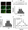
Figure 6. IL-1β overexpression in spinal cord was related to mechanical allodynia in VZV infected rats, and activated astrocytes were the only source of IL-1β.
(A–C′) Double immunofluorescent staining showed that IL-1β-immunoreactivity was localized in GFAP-immunopositive cells but not OX42-immunopositive cells in spinal cord of VZV infected rats. Bars = 20 µm. (D) IL-1β expression was significantly increased in spinal cord of VZV infected rats compared to Naive rats and Mock infected rats. Intrathecal treatment with LAA (astrocytic specific toxin) significantly reduced IL-1β overexpression in VZV infected rats. (E) Using real-time RT-PCR, it was found that the spinal mRNA expression of IL-1β was significantly upregulated in VZV infected rats compared to naive rats and mock infected rats. (F) Intrathecal injection of Pentoxifylline (cytokine inhibitor) or IL-1ra (interleukin-1 receptor antagonist) could significantly attenuated the allodynia. All data were calculated as mean ± SEM (n = 10/group). * P<0.05, ** P<0.01 vs. Naive rats and Mock infected rats; ΔP<0.05 vs. post-infection 2 weeks rats in D and E. * P<0.05, ** P<0.01 vs. saline (vehicle) treated VZV infected rats in F.
Figure 7
Figure 7. IL-1β released from astrocyte induced NMDA receptor phosphorylation in spinal dorsal horn neurons in VZV infected rats, which was related to mechanical allodynia.
(A–C) Double immunofluorescent staining showed that P-NR1-immunoreactivity and IL-1RI-immunoreactivity were totally double-labeled in spinal dorsal horn of VZV infected rats. Bar = 50 µm. (D) The phosphorylation of NR1 was significantly increased in VZV infected rats compared to Naive rats and Mock infected rats. Intrathecal treatment with LAA (astrocytic specific toxin), PF (cytokine inhibitor) or IL-1ra (interleukin-1 receptor antagonist) could significantly reduce the phosphorylation of NR1 in VZV infected rats. (E) Intrathecal injection of AP5 (NMDA receptor antagonist) or MK-801 (non-competitive NMDA receptor antagonist) could significantly attenuated the allodynia. All data were calculated as mean ± SEM (n = 10/group). * P<0.05, ** P<0.01 vs. Naive rats and Mock infected rats; ΔP<0.05 vs. post-infection 2 weeks rats in D. * P<0.05, ** P<0.01 vs. saline (vehicle) treated VZV infected rats in E.

Similar articles

Cited by

References

    1. Dworkin RH, Portenoy RK. Pain and its persistence in herpes zoster. Pain. 1996;67:241–251. - PubMed
    1. Bowsher D. The management of postherpetic neuralgia. Postgrad Med J. 1997;73:623–629. - PMC - PubMed
    1. Lydick E, Epstein RS, Himmelberger D, White CJ. Herpes zoster and quality of life: a self-limited disease with severe impact. Neurology. 1995;45:S52–53. - PubMed
    1. Bowsher D. Pathophysiology of postherpetic neuralgia: towards a rational treatment. Neurology. 1995;45(Suppl. 8):S56–S57. - PubMed
    1. Gilden DH. Herpes zoster with postherpetic neuralgia: persisting pain and frustration. N Engl J Med. 1994;330:932–934. - PubMed

Publication types