

De novo malignant solitary fibrous tumor of the kidney
- PMID: 21970525
- PMCID: PMC3195699
- DOI: 10.1186/1746-1596-6-96
De novo malignant solitary fibrous tumor of the kidney
Abstract
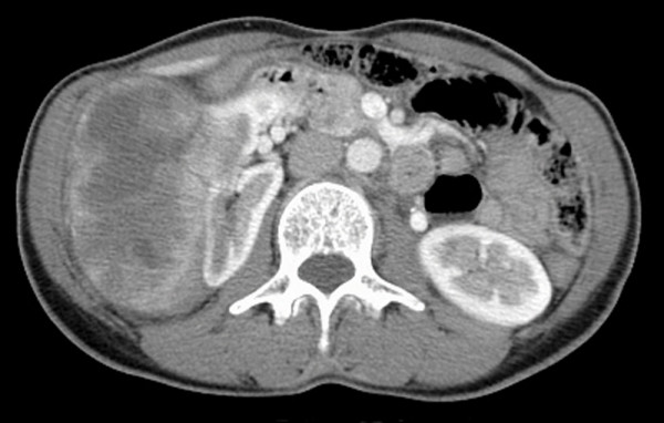
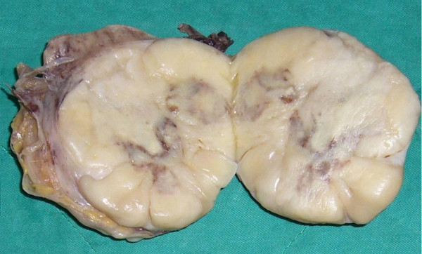
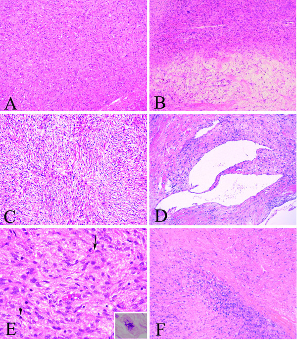
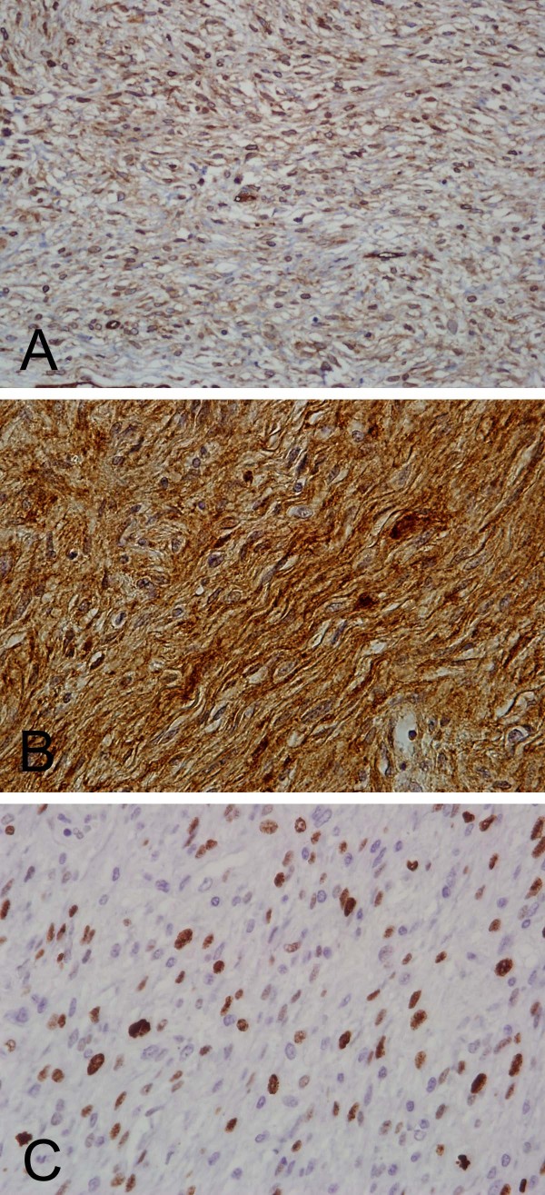
The kidney is a relatively infrequent site for solitary fibrous tumor (SFT). Among the previously reported cases, only two cases of malignant renal SFT developing via dedifferentiation from a pre-existing benign SFT have been reported. Here we reported a case of de novo malignant renal SFT clinically diagnosed as renal cell carcinoma in a 50-year-old woman. The tumor was circumscribed but unencapsulated and showed obvious hemorrhagic necrosis. Microscopically, the tumor was composed of patternless sheets of alternating hypercellular and hypocellular areas of spindle cells displaying mild to moderate nuclear atypia, frequent mitoses up to 8 per 10 high power fields, and a 20% Ki-67 proliferative index. Immunohistochemical studies revealed reactivity for CD34, CD99 and vimentin, with no staining for all other markers, confirming the diagnosis of SFT. No areas of dedifferentiation were seen after extensive sampling. Based on the pathologic and immunohistochemical features, a diagnosis of de novo malignant renal SFT was warranted. Our report expands the spectrum of malignant progression in renal SFTs. Even though this patient has been disease-free for 30 months, long-term follow-up is still mandatory.
Figures
Similar articles
-
A GRIA2 and PAX8-positive renal solitary fibrous tumor with NAB2-STAT6 gene fusion.Diagn Pathol. 2015 Sep 4;10:155. doi: 10.1186/s13000-015-0386-x. Diagn Pathol. 2015. PMID: 26337721 Free PMC article.
-
A huge malignant solitary fibrous tumor of kidney: case report and review of the literature.Diagn Pathol. 2014 Jan 20;9:13. doi: 10.1186/1746-1596-9-13. Diagn Pathol. 2014. PMID: 24443842 Free PMC article. Review.
-
A rare solitary fibrous tumour of kidney.JNMA J Nepal Med Assoc. 2013 Apr-Jun;52(190):388-90. JNMA J Nepal Med Assoc. 2013. PMID: 24362666
-
Intrapulmonary solitary fibrous tumors: clinicopathologic and immunohistochemical study of 24 cases.Am J Surg Pathol. 2013 Feb;37(2):155-66. doi: 10.1097/PAS.0b013e31826a92f5. Am J Surg Pathol. 2013. PMID: 23108019
-
Pediatric renal solitary fibrous tumor: report of a rare case and review of the literature.Int J Surg Pathol. 2015 Feb;23(1):34-47. doi: 10.1177/1066896913492847. Epub 2013 Jul 1. Int J Surg Pathol. 2015. PMID: 23816824 Review.
Cited by
-
Malignant solitary fibrous tumor of the kidney with IGF2 secretion and without hypoglycemia.World J Surg Oncol. 2024 Jul 9;22(1):179. doi: 10.1186/s12957-024-03342-4. World J Surg Oncol. 2024. PMID: 38982409 Free PMC article.
-
Pulmonary sclerosing hemangioma presenting with dense spindle stroma cells: a potential diagnostic pitfall.Diagn Pathol. 2012 Dec 10;7:174. doi: 10.1186/1746-1596-7-174. Diagn Pathol. 2012. PMID: 23227905 Free PMC article.
-
Renal malignant solitary fibrous tumor with single lymph node involvement: report of unusual metastasis and review of the literature.Onco Targets Ther. 2014 May 8;7:679-85. doi: 10.2147/OTT.S51664. eCollection 2014. Onco Targets Ther. 2014. PMID: 24855378 Free PMC article.
-
Collecting duct carcinoma of the kidney: a clinicopathological study of five cases.Diagn Pathol. 2013 Jun 17;8:96. doi: 10.1186/1746-1596-8-96. Diagn Pathol. 2013. Retraction in: Diagn Pathol. 2015 Mar 26;10:15. doi: 10.1186/s13000-015-0239-7. PMID: 23773436 Free PMC article. Retracted.
-
Role of Immunohistochemistry in the Diagnosis of Solitary Fibrous Tumor, a Review.Iran J Pathol. 2016 Summer;11(3):195-203. Iran J Pathol. 2016. PMID: 27799967 Free PMC article. Review.
References
-
- Weiss SW, Goldblum JR. In: Soft Tissue Tumor. 5. Weiss SW, Goldblum JR, editor. Philadephia: Mosby Elsevier; 2008. Soft tissue tumors of intermediate malignancy of uncertain type; pp. 1093–1160.
-
- Guillou LFJ, Fletcher CDM, Mandahi N. In: World Health Organization Classification of Tumours: Pathology and Genetics of Tumours of Soft Tissue and Bone. Fletcher CDM, Unni KK, Mertens F, editor. Lyon: IARCPress; 2002. Extrapleural solitary fibrous tumour and hemangiopericytoma; pp. 86–90.
Publication types
MeSH terms
Substances
LinkOut - more resources
Full Text Sources
Medical
