. 2012 Mar 20;29(5):735-46.
doi: 10.1089/neu.2011.2008. Epub 2011 Dec 8.

Quantitative CT improves outcome prediction in acute traumatic brain injury

Affiliations

Quantitative CT improves outcome prediction in acute traumatic brain injury

Esther L Yuh et al. J Neurotrauma. .

Abstract

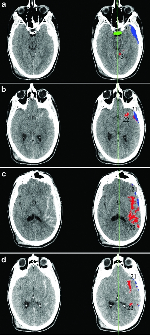
The admission noncontrast head computed tomography (CT) scan has been demonstrated to be one of several key early clinical and imaging features in the challenging problem of prediction of long-term outcome after acute traumatic brain injury (TBI). In this study, we employ two novel approaches to the problem of imaging classification and outcome prediction in acute TBI. First, we employ the novel technique of quantitative CT (qCT) image analysis to provide more objective, reproducible measures of the abnormal features of the admission head CT in acute TBI. We show that the incorporation of quantitative, rather than qualitative, CT features results in a significant improvement in prediction of the 6-month Extended Glasgow Outcome Scale (GOS-E) score over a wide spectrum of injury severity. Second, we employ principal components analysis (PCA) to demonstrate the interdependence of certain predictive variables. Relatively few prior studies of outcome prediction in acute TBI have used a multivariate approach that explicitly takes into account the potential covariance among clinical and CT predictive variables. We demonstrate that several predictors, including midline shift, cistern effacement, subdural hematoma volume, and Glasgow Coma Scale (GCS) score are related to one another. Rather than being independent features, their importance may be related to their status as surrogate measures of a more fundamental underlying clinical feature, such as the severity of intracranial mass effect. We believe that objective computational tools and data-driven analytical methods hold great promise for neurotrauma research, and may ultimately have a role in image analysis for clinical care.


Figures

FIG. 1.
FIG. 1.
Software estimation of volumes of acute intracranial hemorrhage. Suspected extra-axial hemorrhagic collections (a–d) are identified through computer analysis of images and assigned a numeric identifier. Most of these initially-identified collections are excluded through further quantitative analysis of their three-dimensional properties. Collections that satisfy criteria for acute intracranial hemorrhage are identified as color overlays (red, subarachnoid hemorrhage; blue, subdural hematoma) on the original computed tomography (CT) images, and the volume of each collection is calculated. Midline shift is measured as the shift of the lateral ventricles relative to the computer-estimated falx plane (green line). The dorsum sella is detected (a, horizontal green bar), and the basal cistern volume is measured as the volume of fluid surrounding and superior to the dorsum sella.
FIG. 2.
FIG. 2.
Distribution of (a) ages and (b) admission Glasgow Coma Scale (GCS) scores in the study population. Both patient age and admission GCS scores demonstrated significant variability. Although slightly over half of the admission GCS scores were in the mild head injury range (GCS 13–15), significant numbers of cases in the moderate and severe head injury categories were included. Principal component analysis (PCA) is based partly on the recognition that predictors without substantial variability are less likely to account for differences in the observed outcome measure.
FIG. 3.
FIG. 3.
Distribution of qualitative computed tomography (CT) features in the study population. (a) Slightly over half of patients had subdural hematoma. (b) Subarachnoid hemorrhage (SAH) and/or intraparenchymal hemorrhage (IPH) was seen in over two-thirds of patients. (c) Epidural hematoma occurred in less than 10% of cases. (d) Slightly over 10% of patients had midline shift exceeding 5 mm. (e) Approximately 25% of patients had some basal cistern effacement, divided evenly between partial and complete effacement.
FIG. 4.
FIG. 4.
Distribution of quantitative computed tomography (qCT) features in the study population. Computer-aided quantitative estimates of (a) subdural hematoma (SDH) volume, (b) subarachnoid hemorrhage (SAH) and intraparenchymal hemorrhage (IPH) volume, (c) epidural hematoma volume, and (d) midline shift. Subdural hematoma was present in slightly over half of patients, with volumes ranging from trace amounts to nearly 40 cubic centimeters. Subarachnoid and intraparenchymal hemorrhage was present in over 70% of patients, usually in small quantities less than 3 cubic centimeters, although volumes of 12 or more cubic centimeters were also observed. Epidural hematoma was observed in less than 10% of patients. Midline shift exceeding 5 mm was seen in slightly more than 10% of patients. Shifts from 7 mm to over 1.5 cm were observed.
FIG. 5.
FIG. 5.
Extended Glasgow Outcome Scale (GOS-E) scores at 6 months following the traumatic brain injury (TBI) event. GOS-E scores at 6 months following head injury also demonstrated wide variability. High variability in the outcome measure in a given study population increases the likelihood of achieving statistical significance of the regression model based on the study population.

References

    1. Chestnut R.M. Ghajar J. Maas A.I. Marion D.W. Servadei F. Teasdale G.M. Unterberg A. von Holst H. Walters B.C. Management and prognosis of severe traumatic brain injury. Part 2: Early indicators of prognosis in severe traumatic brain injury. J. Neurotrauma. 2000;17:557–627.
    1. Chieregato A. Fainardi E. Morselli-Labate A.M. Antonelli V. Compagnone C. Targa L. Kraus J. Servadei F. Factors associated with neurological outcome and lesion progression in traumatic subarachnoid hemorrhage patients. Neurosurgery. 2005;56:671–680. - PubMed
    1. Chun K.A. Manley G.T. Stiver S.I. Aiken A.H. Phan N. Wang V. Meeker M. Cheng S.C. Gean A.D. Wintermark M. Interobserver variability in the assessment of CT imaging features of traumatic brain injury. J Neurotrauma. 2010;27:325–330. - PMC - PubMed
    1. Eisenberg H.M. Gary H.E., Jr. Aldrich E.F. Saydjari C. Turner B. Foulkes M.A. Jane J.A. Marmarou A. Marshall L.F. Young H.F. Initial CT findings in 753 patients with severe head injury: A report from the NIH Traumatic Coma Data Bank. J. Neurosurg. 1990;73:688–698. - PubMed
    1. Hukkelhoven C.W. Steyerberg E.W. Habbema J.D. Farace E. Marmarou A. Murray G.D. Marshall L.F. Maas A.I. Predicting outcome after traumatic brain injury: Development and validation of a prognostic score based on admission characteristics. J. Neurotrauma. 2005;22:1025–1039. - PubMed

Publication types