. 2011 Dec 23;286(51):43972-43983.
doi: 10.1074/jbc.M111.301093. Epub 2011 Oct 4.

Autotaxin regulates vascular development via multiple lysophosphatidic acid (LPA) receptors in zebrafish

Affiliations

Autotaxin regulates vascular development via multiple lysophosphatidic acid (LPA) receptors in zebrafish

Hiroshi Yukiura et al. J Biol Chem. .

Abstract

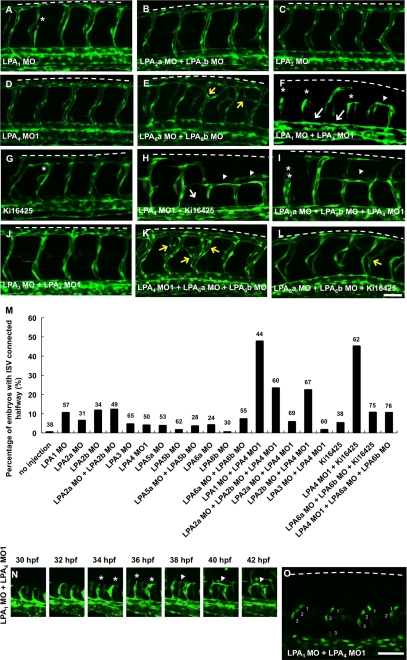
Autotaxin (ATX) is a multifunctional ecto-type phosphodiesterase that converts lysophospholipids, such as lysophosphatidylcholine, to lysophosphatidic acid (LPA) by its lysophospholipase D activity. LPA is a lipid mediator with diverse biological functions, most of which are mediated by G protein-coupled receptors specific to LPA (LPA1-6). Recent studies on ATX knock-out mice revealed that ATX has an essential role in embryonic blood vessel formation. However, the underlying molecular mechanisms remain to be solved. A data base search revealed that ATX and LPA receptors are conserved in wide range of vertebrates from fishes to mammals. Here we analyzed zebrafish ATX (zATX) and LPA receptors both biochemically and functionally. zATX, like mammalian ATX, showed lysophospholipase D activity to produce LPA. In addition, all zebrafish LPA receptors except for LPA5a and LPA5b were found to respond to LPA. Knockdown of zATX in zebrafish embryos by injecting morpholino antisense oligonucleotides (MOs) specific to zATX caused abnormal blood vessel formation, which has not been observed in other morphant embryos or mutants with vascular defects reported previously. In ATX morphant embryos, the segmental arteries sprouted normally from the dorsal aorta but stalled in midcourse, resulting in aberrant vascular connection around the horizontal myoseptum. Similar vascular defects were not observed in embryos in which each single LPA receptor was attenuated by using MOs. Interestingly, similar vascular defects were observed when both LPA1 and LPA4 functions were attenuated by using MOs and/or a selective LPA receptor antagonist, Ki16425. These results demonstrate that the ATX-LPA-LPAR axis is a critical regulator of embryonic vascular development that is conserved in vertebrates.


Figures

FIGURE 1.
FIGURE 1.
Zebrafish ATX is a potent LPA-producing enzyme. A, domain structures of human and zebrafish ATXs. The amino acid identity of each domain is indicated. B and C, lysophospholipase D activity of recombinant zebrafish ATX. HEK293 cells were transfected with plasmid DNA encoding C-terminally Myc-tagged wild-type (zATX-myc-pCAGGS) and mutant ATX (zATX (T205A)-myc-pCAGGS). Secreted ATX in the conditioned medium (B) was analyzed by Western blotting using anti-Myc monoclonal antibody (9E10). Lysophospholipase D activity of wild-type (Wt) and mutant zebrafish ATX (T205A) (C) was analyzed using 1-myristoyl lysophosphatidylcholine as a substrate. D, nucleotide phosphodiesterase activity of mouse and zebrafish ATX, using p-nitrophenol-TMP as a substrate. E and F, phosphodiesterase activities of zebrafish ATX against LPC (E) and pNP-TMP (F) were examined with the indicated concentrations of substrates. Inset, Lineweaver-Burk plot to calculate Km and Vmax values. Error bars, S.D.
FIGURE 2.
FIGURE 2.
Substrate specificity of ATX is conserved between mouse and zebrafish. A, effects of the polar headgroup of lysophospholipids on the enzymatic activity of ATX. 1-oleoyl-LPE, -LPS, -LPI, and -LPC were incubated at 37 °C for 3 h in the presence of zebrafish or mouse ATX. The amount of LPA produced was quantified by an enzyme-linked colorimetric method. B, substrate specificity of zebrafish and mouse ATX for LPC with a variety of fatty acids and SPC. LPC- and SPC-hydrolyzing activities were measured in the presence of 0.05% Triton X-100. C, catalytic activity of mouse and zebrafish ATX against physiological substrates. Recombinant mouse and zebrafish ATX were incubated in ATX-depleted plasma prepared using anti-mouse ATX monoclonal antibodies. The amount of LPA produced by recombinant ATX was measured by mass spectrometry. Error bars indicate the S.D. of the mean.
FIGURE 3.
FIGURE 3.
Zebrafish ATX stimulates the cell motility. Cell motility-stimulating activities of recombinant zebrafish ATX. Zebrafish ATX recombinant protein promoted the motility of MDA-MB-231 cells in a Ki16425-sensitive manner. Error bars, S.D.
FIGURE 4.
FIGURE 4.
Expression pattern of zebrafish ATX gene at stage of development. A, ATX mRNA expression at the indicated times postfertilization, as measured by quantitative RT-PCR. Values are normalized to the expression level of elfa1 mRNA. Error bars indicate the S.D. of the mean. B and C, whole mount in situ hybridization analyses with antisense (B and B′) and sense (C and C′) probes of atx at 30 hpf. ATX mRNA is expressed in the head region (arrow), the neural tube (N), and the vasculature (V) including the DA and the PCV at 30 hpf. Lateral views, anterior to the left. Scale bar, 50 μm.
FIGURE 5.
FIGURE 5.
Zebrafish ATX is important for the segmental artery development. A, Western blot analysis using an antibody against zebrafish ATX. Embryos were injected with the indicated MO. Embryo lysates were prepared at 48 hpf. Tubulin expression was used as a loading control. B and C, effects of ATX morpholino. As compared with uninjected embryos (B), embryos injected with ATX MO1 (C) caused edema in heart and head regions (arrows). D–F, confocal images of fli1:EGFP embryos at 48 hpf. Lateral views, anterior to the left. In embryos injected with control MO (D), the SAs extended from the DA and formed the DLAVs. In contrast, in ATX MO1-injected embryos (E) and ATX MO2-injected embryos (F), the SAs stalled around the horizontal myoseptum (asterisk) and aberrantly connected to the neighboring ISV (arrowheads). Dotted lines, dorsal edge of the embryos. G, graph representing the percentage of embryos with aberrant ISV connection around the horizontal myoseptum at 48 hpf (n = 31–35; ***, p < 0.001 by χ2 test). H and I, confocal time lapse analysis of fli1:EGFP embryos from 30 to 42 hpf. Control embryos show the normal SA sprouts (H). By contrast, in ATX morphant embryos, the ISV initially stalled around the horizontal myoseptum at 34 hpf (asterisk) and connected the neighboring ISV abnormally at 42 hpf (arrowhead) (I). J and K, confocal images of fli1:nEGFP embryos at 32 hpf. Each SA consists of two or three endothelial cells both in control (J) and ATX morphant embryos (K). Scale bar, 50 μm.
FIGURE 6.
FIGURE 6.
Responses of LPA receptor homologues in zebrafish. Each LPA receptor was transfected into HEK293 cells, and the amount of TGF-α released upon LPA stimulation was measured. The LPA response was observed in all LPA receptor homologues except for LPA5a or LPA5b. Error bars indicate the S.D. of the mean.
FIGURE 7.
FIGURE 7.
Inhibitory effects of Ki16425 on the activation of LPA receptor homologues in zebrafish. Ki16425 inhibited the activation of LPA1, LPA2a, LPA2b, and LPA3 but not the activation LPA4, LPA6a, or LPA6b upon LPA stimuli.
FIGURE 8.
FIGURE 8.
LPA1 and LPA4 are important for segmental artery development. Confocal images of fli1:EGFP embryos at 48 hpf. Lateral views, anterior to the left. Embryos were treated with MOs or 120 μm Ki16425 dissolved in 1% DMSO. Embryos treated with LPA1 (A), LPA2a/LPA2b (B), LPA3 (C), LPA4 (D), LPA3/LPA4 (J) MOs and Ki16425 (G) showed almost normal ISV sprouts, except for a few SAs that stalled around the horizontal myoseptum (asterisks) in embryos treated with LPA1 MO and Ki16425. Embryos treated with LPA1/LPA4 (F) and LPA2a/LPA2b/LPA4 (I) MOs and LPA4 MO with Ki16425 (H) displayed the vascular phenotype of abnormal connection between neighboring ISVs (arrowheads in F, H, and I). Embryos treated with LPA6a/LPA6b (E) and LPA4/LPA6a/LPA6b (K) MOs and LPA6a/LPA6b MOs with Ki16425 (L) showed ISV branching (yellow arrows). Embryos treated with LPA1/LPA4 MOs (F) and LPA4 MO with Ki16425 (H) also showed the dissociation between SA and DA (arrows). M, percentage of embryos with aberrant ISV connections around the horizontal myoseptum at 48 hpf (n = 24–76). N, confocal time lapse analysis of fli1:EGFP embryos injected with LPA1/LPA4 MOs from 30 to 42 hpf. ISVs initially stalled around the horizontal myoseptum at 34 hpf (asterisk) and connected to the neighboring ISVs abnormally at 42 hpf (arrowhead). O, confocal images of fli1:nEGFP embryos injected with LPA1/LPA4 MOs at 32 hpf. Each SA consists of two or three endothelial cells. Scale bar, 50 μm. Numbers 1–3 indicate the number of endothelial cells in one ISV.

References

    1. Stracke M. L., Krutzsch H. C., Unsworth E. J., Arestad A., Cioce V., Schiffmann E., Liotta L. A. (1992) J. Biol. Chem. 267, 2524–2529 - PubMed
    1. Umezu-Goto M., Kishi Y., Taira A., Hama K., Dohmae N., Takio K., Yamori T., Mills G. B., Inoue K., Aoki J., Arai H. (2002) J. Cell Biol. 158, 227–233 - PMC - PubMed
    1. Tokumura A., Majima E., Kariya Y., Tominaga K., Kogure K., Yasuda K., Fukuzawa K. (2002) J. Biol. Chem. 277, 39436–39442 - PubMed
    1. Aoki J. (2004) Semin. Cell Dev. Biol. 15, 477–489 - PubMed
    1. Tanaka M., Okudaira S., Kishi Y., Ohkawa R., Iseki S., Ota M., Noji S., Yatomi Y., Aoki J., Arai H. (2006) J. Biol. Chem. 281, 25822–25830 - PubMed

Publication types

Substances