

Cerebral microvascular inflammation in DOCA salt-induced hypertension: role of angiotensin II and mitochondrial superoxide
- PMID: 21971354
- PMCID: PMC3272604
- DOI: 10.1038/jcbfm.2011.139
Cerebral microvascular inflammation in DOCA salt-induced hypertension: role of angiotensin II and mitochondrial superoxide
Abstract
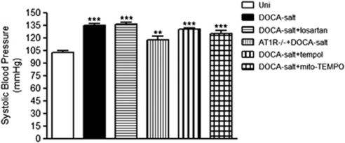
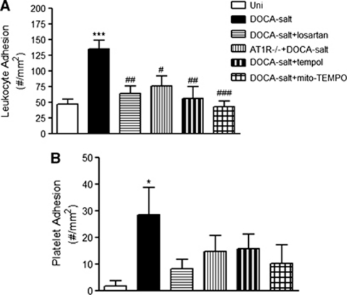
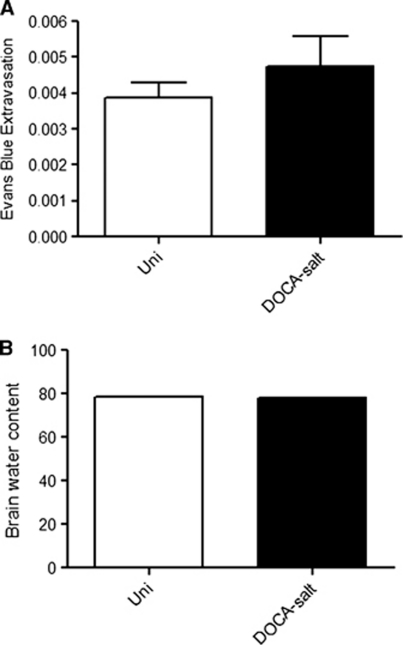
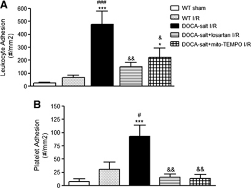
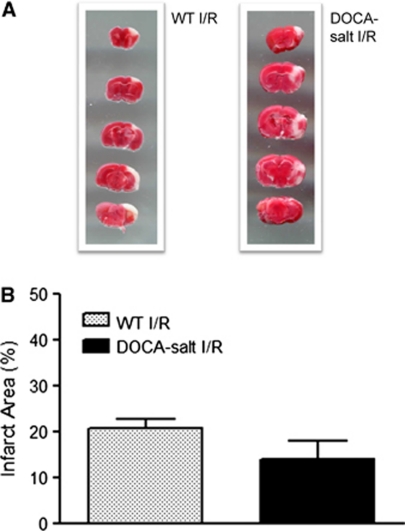
Angiotensin II-mediated hypertension (HTN) is accompanied by a pro-inflammatory and pro-thrombotic state in the cerebral microvasculature. Whether comparable phenotypic changes are elicited in other models of HTN remains unclear. Using wild-type mice with deoxycorticosterone acetate (DOCA) salt-induced HTN and intravital microscopy, we observed significant increases in the adhesion of both leukocytes and platelets in cerebral venules, compared with uninephrectomized control mice, without an accompanying increase in blood-brain barrier permeability. The cell-cell interactions in hypertensive mice were more pronounced after ischemic stroke, but no difference in infarct size was detected. The blood cell recruitment was largely prevented in the following groups of DOCA salt mice: losartan (angiotensin II AT1 receptor blocker) treated, AT1 receptor knockout mice, tempol (a membrane-permeable oxygen radical scavenger) treated, and mito-TEMPO (a mitochondria-targeted antioxidant) treated. A similar pattern of protection was noted in mice subjected to ischemic stroke. The blunted cell recruitment responses were not accompanied by reductions in blood pressure (BP). These findings implicate mitochondria-derived oxygen radicals and angiotensin II in the cerebral inflammation associated with DOCA salt HTN and suggests that BP per se is not a critical determinant of the phenotypic changes that accompany HTN, even after ischemic stroke.
Figures
References
-
- Arumugam TV, Salter JW, Chidlow JH, Ballantyne CM, Kevil CG, Granger DN. Contributions of LFA-1 and Mac-1 to brain injury and microvascular dysfunction induced by transient middle cerebral artery occlusion. Am J Physiol Heart Circ Physiol. 2004;287:H2555–H2560. - PubMed
-
- Basso N, Ruiz P, Mangiarua E, Taquini AC.1981Renin-like activity in the rat brain during the development of DOC-salt hypertension Hypertension 3(Suppl II): II14–II7. - PubMed
-
- Bird JE, Moreland S, Waldron TL, Powell JR. Antihypertensive effects of a novel endothelin-A receptor antagonist in rats. Hypertension. 1995;25:1191–1195. - PubMed
-
- Coyle P, Jokelainen PT. Differential outcome to middle cerebral artery occlusion in spontaneously hypertensive stroke-prone rats (SHRSP) and Wistar Kyoto (WKY) rats. Stroke. 1983;14:605–611. - PubMed
-
- Crofton JT, Share L, Shade RE, Lee-Kwon WJ, Manning M, Sawyer WH. The importance of vasopressin in the development and maintenance of DOC-salt hypertension in the rat. Hypertension. 1979;1:31–38. - PubMed
Publication types
MeSH terms
Substances
Grants and funding
LinkOut - more resources
Full Text Sources
Other Literature Sources
Medical
Research Materials
