

Review
doi: 10.3233/JAD-2011-0007.
DTI analyses and clinical applications in Alzheimer's disease
Affiliations
- PMID: 21971468
- PMCID: PMC3294372
- DOI: 10.3233/JAD-2011-0007
Item in Clipboard
Review
DTI analyses and clinical applications in Alzheimer's disease
J Alzheimers Dis.
2011.
Abstract
DTI is one of the most effective MR tools for the investigation of the brain anatomy. In addition to the gray matter, histopathological studies indicate that white matter is also a good target for both the early diagnosis of AD and for monitoring disease progression, which motivates us to use DTI to study AD patients in vivo. There are already a large amount of studies reporting significant differences between AD patients and controls, as well as to predict progression of disease in symptomatic non-demented individuals. Application of these findings in clinical practice remains to be demonstrated.
Figures

Comparison of conventional T1- (A) and T2- (B) weighted images, and DTI-derived mean diffusivity (MD) (C), fractional anisotropy (FA) (D), and color-coded orientation (E) maps of cognitively normal 72-year-old woman (upper row) and 70-year-old woman with Alzheimer’s disease. The areas surrounded by yellow rectangles in (E) are magnified and shown in (F) [left (F-1): from the cognitively normal woman; right (F-2): the Alzheimer’s disease patient]. The yellow arrows indicate the cingulum hippocampal part.
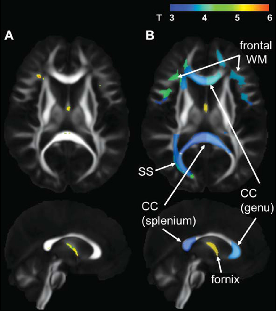
Statistical group comparison of FA after image normalization. Nineteen patients with Alzheimer’s disease and 22 age-matched cognitively normal participants were compared, and the areas with significance (t-test, p < 0.05 after correction for multiple comparisons using a false discovery rate) were shown with color scaling overlaid on the averaged FA map (A): Results from voxel-based analysis. (B): Results from atlas-based analysis. See Fig. 3. for the parcellation map used in this atlas-based analysis. CC, corpus callosum; SS, sagittal stratum; WM, white matter.

Atlas-based analysis. The original FA map from a patient with Alzheimer’s disease (A) was normalized to the atlas space (B). The atlas used as the template is shown in (C). After image normalization, pre-defined three-dimensional ROIs (color-contours, called a parcellation map) in the atlas space enables researchers to measure the FA value of each parcel. This parcellation map can be inversely transformed to the original space to measure the volume of each parcel, as well as to measure the FA value of each parcel without normalization-related artifacts (eg., effects of interpolation after normalization).
References
-
- Beaulieu C, Allen PS. Determinants of anisotropic water diffusion in nerves. Magn Reson Med. 1994;31:394–400. - PubMed
-
- Moseley ME, Cohen Y, Kucharczyk J, Mintorovitch J, Asgari HS, Wendland MF, Tsuruda J, Norman D. Diffusion-weighted MR imaging of anisotropic water diffusion in cat central nervous system. Radiology. 1990;176:439–445. - PubMed
-
- Pierpaoli C, Basser PJ. Toward a quantitative assessment of diffusion anisotropy. Magn Reson Med. 1996;36:893–906. - PubMed
-
- Pierpaoli C, Jezzard P, Basser PJ, Barnett A, Di Chiro G. Diffusion tensor MR imaging of human brain. Radiology. 1996;201:637–648. - PubMed
-
- Makris N, Worth AJ, Sorensen AG, Papadimitriou GM, Wu O, Reese TG, Wedeen VJ, Davis TL, Stakes JW, Caviness VS, Kaplan E, Rosen BR, Pandya DN, Kennedy DN. Morphometry of in vivo human white matter association pathways with diffusion-weighted magnetic resonance imaging. Ann Neurol. 1997;42:951–962. - PubMed
Publication types
MeSH terms
Grants and funding
LinkOut - more resources
Full Text Sources
Medical
