

Microarray analysis reveals age-related differences in gene expression during the development of osteoarthritis in mice
- PMID: 21972019
- PMCID: PMC3269534
- DOI: 10.1002/art.33388
Microarray analysis reveals age-related differences in gene expression during the development of osteoarthritis in mice
Abstract
Objective: To better understand the contribution of age to the development of osteoarthritis (OA).
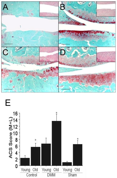
Methods: Surgical destabilization of the medial meniscus (DMM) was used to model OA in 12-week-old and 12-month-old male C57BL/6 mice. OA severity was evaluated histologically. RNA used for microarray and real-time polymerase chain reaction analysis was isolated from joint tissue collected from the medial side of the joint, including cartilage, meniscus, subchondral bone, and the joint capsule with synovium. Computational analysis was used to identify patterns of gene expression, and immunohistochemistry was used to evaluate tissue distribution of selected proteins.
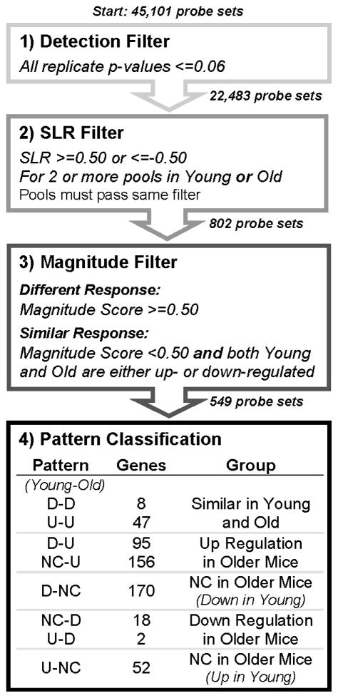
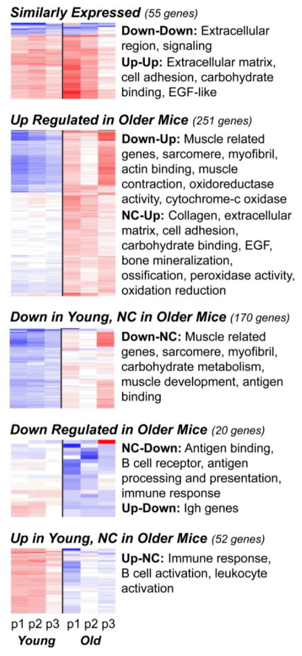
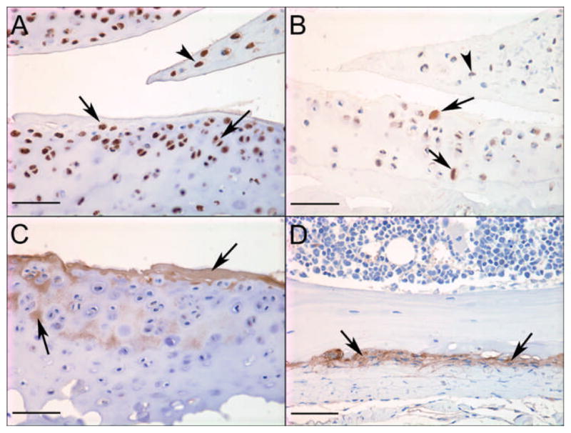
Results: OA was more severe in older mice than in young mice. Only 55 genes showed a similar expression with DMM-induced OA in the 2 age groups, while 493 genes showed differential expression, the majority having increased expression in older mice. Functional categories for similarly expressed genes included extracellular matrix- and cell adhesion-related genes; differentially expressed genes included those related to muscle structure and development and immune response genes. Comparison of expression in sham-operated control joints revealed an age-related decrease in matrix gene expression and an increase in immune and defense response gene expression. Interleukin-33 was present in multiple joint tissue cells, while CCL21 was more localized to chondrocytes and meniscal cells. Periostin was found in the extracellular matrix of cartilage and meniscus.
Conclusion: Age affects both the basal pattern of gene expression in joint tissues and the response to surgically induced OA. Examining tissue from the joint beyond only cartilage revealed novel genes and proteins that would be important to consider in OA.
Copyright © 2012 by the American College of Rheumatology.
Figures
References
-
- Blagojevic M, Jinks C, Jeffery A, Jordan KP. Risk factors for onset of osteoarthritis of the knee in older adults: a systematic review and meta-analysis. Osteoarthritis Cartilage. 2010;18:24–33. - PubMed
-
- Roos H, Adalberth T, Dahlberg L, Lohmander LS. Osteoarthritis of the knee after injury to the anterior cruciate ligament or meniscus: the influence of time and age. Osteoarthritis Cartilage. 1995;3:261–7. - PubMed
-
- Aigner T, Zien A, Gehrsitz A, Gebhard PM, McKenna L. Anabolic and catabolic gene expression pattern analysis in normal versus osteoarthritic cartilage using complementary DNA-array technology. Arthritis Rheum. 2001;44:2777–89. - PubMed
-
- Aigner T, Fundel K, Saas J, Gebhard PM, Haag J, Weiss T, et al. Large-scale gene expression profiling reveals major pathogenetic pathways of cartilage degeneration in osteoarthritis. Arthritis Rheum. 2006;54:3533–44. - PubMed
Publication types
MeSH terms
Substances
Grants and funding
LinkOut - more resources
Full Text Sources
Other Literature Sources
Medical
