. 2011 Mar;32(3):299-308.
doi: 10.1002/humu.21426.

Nonsense mutation-associated Becker muscular dystrophy: interplay between exon definition and splicing regulatory elements within the DMD gene

Collaborators, Affiliations

Nonsense mutation-associated Becker muscular dystrophy: interplay between exon definition and splicing regulatory elements within the DMD gene

Kevin M Flanigan et al. Hum Mutat. 2011 Mar.

Abstract

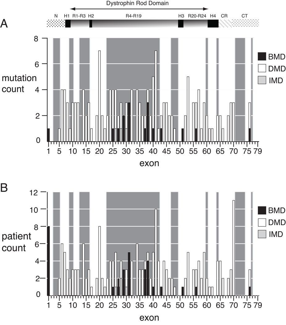
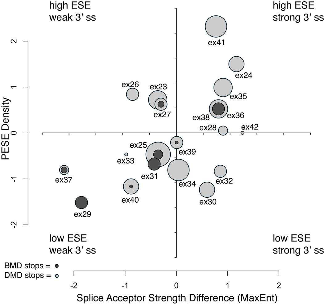
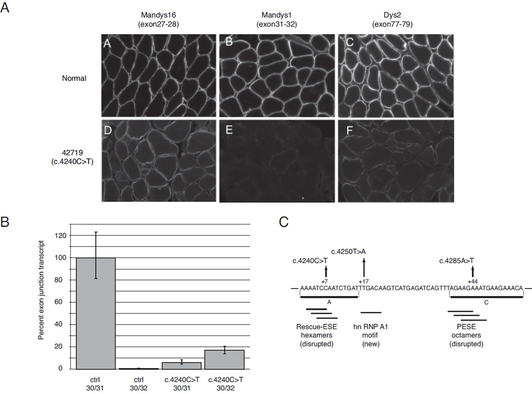
Nonsense mutations are usually predicted to function as null alleles due to premature termination of protein translation. However, nonsense mutations in the DMD gene, encoding the dystrophin protein, have been associated with both the severe Duchenne Muscular Dystrophy (DMD) and milder Becker Muscular Dystrophy (BMD) phenotypes. In a large survey, we identified 243 unique nonsense mutations in the DMD gene, and for 210 of these we could establish definitive phenotypes. We analyzed the reading frame predicted by exons flanking those in which nonsense mutations were found, and present evidence that nonsense mutations resulting in BMD likely do so by inducing exon skipping, confirming that exonic point mutations affecting exon definition have played a significant role in determining phenotype. We present a new model based on the combination of exon definition and intronic splicing regulatory elements for the selective association of BMD nonsense mutations with a subset of DMD exons prone to mutation-induced exon skipping.


Figures

Figure 1
Figure 1
Exon distribution of dystrophin nonsense mutations by phenotype. A) The number of mutations per exon for unique mutation sites (N=161) is shown for Duchenne (open boxes), intermediate (light boxes), and Becker (dark boxes) phenotypes. Exons with in-frame flanking exon context are highlighted by the light grey background shading, and for each exon within the shaded region, single exon skipping will result in bypass of the truncating mutation. Only a single mutation site (c.3500) is counted twice, due to a discrepant phenotype between kindreds, as discussed in the text. B) The number of mutations per exon by patient (N=210). The exon location of dystrophin protein domains are shown above the graphs: NH2 terminus (N), rod domain spectrin repeats 1 through 24 (R), rod domain hinges 1 through 4 (H), cysteine-rich domain (CR), carboxyl terminus (CT).
Figure 2
Figure 2
Exonic splicing enhancer density versus relative splice acceptor site strength for in-frame exons 23 through 42. Normalized scores (mean = 0, variance = 1) are plotted for PESE density and the relative 3’ss strength calculated as the difference between 3’ss scores of the mutation-containing exon and the next distal 3’ss. The circles represent individual exons, where the radius is equal to the total number of mutation sites observed for that exon. The nested ratio of BMD (dark gray) versus DMD (light gray) mutations is indicated for each exon.
Figure 3
Figure 3
The positions of BMD, DMD and IMD nonsense mutations for in-frame exons 23 through 42. The exon locations of BMD mutations are indicated by dark gray squares, DMD mutations by light gray squares and IMD mutations by medium gray squares. Creation or ablation of an ESE hexamer is indicated by an up or down arrow, respectively, above the mutation site, while creation or ablation of an ESS site by an up or down arrow below the site. The ESE and ESS hexamer set were the merged lists of PESE, Rescue-ESE, PESS and FAS-hex3 sequences previously described (Ke, et al., 2008). Overlap of mutations between this study and other studies (see Supp. Table S1 for list of mutations shown) is shown within each rectangle by no symbol (this study only), filled circle (this study and other studies), and an open circle (other studies only). Exon sizes are drawn to scale and flanking intron sizes are indicated in base pairs.
Figure 4
Figure 4
RNA and protein level analysis of exon 31 skipping in BMD patient 42719 (c.4240C>T). A) Immunofluorescent analysis of tissue sections with dystrophin antibodies with epitopes encoded by exons 27–28 for Mandys16, exons 31–32 for Mandys1, and exons 77–79 for Dys2. Positive staining indicates presence of the amino acids encoded in epitope-containing exons in the synthesized dystrophin protein; images represent projections of z-series stacks of confocal sections. B) Comparative CT (ΔΔ CT) analysis of exon 30–31 junction and exon 30–32 junction levels from total RNA extracted from muscle sections. C) Schematic diagram of exon 31 shows the location of BMD mutations: p.Gln1414X (c.4240C>T), p.Leu1417X (c.4250T>A), and p.Lys1429X (c.4285A>T). Coordinates are from the intron 30 – exon 31 junction, and horizontal lines below the sequence show predicted ESE/ESS motifs. Horizontal lines A and C are putative ESE regions predicted by Disset et al. (Disset, et al., 2006).

References

    1. Aartsma-Rus A, van Ommen GJ. Antisense-mediated exon skipping: a versatile tool with therapeutic and research applications. RNA. 2007;13(10):1609–1624. - PMC - PubMed
    1. Barash Y, Calarco JA, Gao W, Pan Q, Wang X, Shai O, Blencowe BJ, Frey BJ. Deciphering the splicing code. Nature. 2010;465(7294):53–59. - PubMed
    1. Brudno M, Gelfand MS, Spengler S, Zorn M, Dubchak I, Conboy JG. Computational analysis of candidate intron regulatory elements for tissue-specific alternative pre-mRNA splicing. Nucleic Acids Res. 2001;29(11):2338–2348. - PMC - PubMed
    1. Cartegni L, Krainer AR. Disruption of an SF2/ASF-dependent exonic splicing enhancer in SMN2 causes spinal muscular atrophy in the absence of SMN1. Nat Genet. 2002;30(4):377–384. - PubMed
    1. Dent KM, Dunn DM, von Niederhausern AC, Aoyagi AT, Kerr L, Bromberg MB, Hart KJ, Tuohy T, White S, den Dunnen JT and others. Improved molecular diagnosis of dystrophinopathies in an unselected clinical cohort. Am J Med Genet A. 2005;134(3):295–298. - PubMed

Publication types