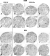
Decreased cytochrome c oxidase subunit VIIa in aged rat heart mitochondria: immunocytochemistry
- PMID: 21972221
- PMCID: PMC3744322
- DOI: 10.1002/ar.21486
Decreased cytochrome c oxidase subunit VIIa in aged rat heart mitochondria: immunocytochemistry
Abstract
Aging decreases oxidative phosphorylation through cytochrome oxidase (COX) in cardiac interfibrillar mitochondria (IFM) in 24-month old (aged) rats compared to 6-month old adult Fischer 344 rats, whereas subsarcolemmal mitochondria (SSM) located beneath the plasma membrane remain unaffected. Immunoelectron microscopy (IEM) reveals in aged rats a 25% reduction in cardiac COX subunit VIIa in cardiac IFM, but not in SSM. In contrast, the content of subunit IV remains unchanged in both SSM and IFM, irrespective of age. These subunits are localized mainly on cristae membranes. In contrast, semi-quantitative immunoblotting, which detects denatured protein, indicates that the content of COX VIIa is similar in IFM and SSM from both aged and adult hearts. IEM provides a sensitive method for precise localizing and quantifying specific mitochondrial proteins. The lack of immunoreaction of COX VIIa subunit by IEM in aged IFM is not explained by a reduction in protein, but rather by a masking phenomenon or by an in situ change in protein structure affecting COX activity.
Copyright © 2011 Wiley-Liss, Inc.
Figures


Similar articles
-
Aging selectively decreases oxidative capacity in rat heart interfibrillar mitochondria.Arch Biochem Biophys. 1999 Dec 15;372(2):399-407. doi: 10.1006/abbi.1999.1508. Arch Biochem Biophys. 1999. PMID: 10600182
-
Interfibrillar cardiac mitochondrial comples III defects in the aging rat heart.Biogerontology. 2002;3(1-2):41-4. doi: 10.1023/a:1015251212039. Biogerontology. 2002. PMID: 12014840
-
Aging decreases electron transport complex III activity in heart interfibrillar mitochondria by alteration of the cytochrome c binding site.J Mol Cell Cardiol. 2001 Jan;33(1):37-47. doi: 10.1006/jmcc.2000.1273. J Mol Cell Cardiol. 2001. PMID: 11133221
-
Ischemia-reperfusion injury in the aged heart: role of mitochondria.Arch Biochem Biophys. 2003 Dec 15;420(2):287-97. doi: 10.1016/j.abb.2003.09.046. Arch Biochem Biophys. 2003. PMID: 14654068 Review.
-
The subunit composition and function of mammalian cytochrome c oxidase.Mitochondrion. 2015 Sep;24:64-76. doi: 10.1016/j.mito.2015.07.002. Epub 2015 Jul 17. Mitochondrion. 2015. PMID: 26190566 Review.
Cited by
-
P23H opsin knock-in mice reveal a novel step in retinal rod disc morphogenesis.Hum Mol Genet. 2014 Apr 1;23(7):1723-41. doi: 10.1093/hmg/ddt561. Epub 2013 Nov 7. Hum Mol Genet. 2014. PMID: 24214395 Free PMC article.
-
Metabolic Complications in Cardiac Aging.Front Physiol. 2021 Apr 29;12:669497. doi: 10.3389/fphys.2021.669497. eCollection 2021. Front Physiol. 2021. PMID: 33995129 Free PMC article. Review.
-
The inhibition of TDP-43 mitochondrial localization blocks its neuronal toxicity.Nat Med. 2016 Aug;22(8):869-78. doi: 10.1038/nm.4130. Epub 2016 Jun 27. Nat Med. 2016. PMID: 27348499 Free PMC article.
-
Can proteomics yield insight into aging aorta?Proteomics Clin Appl. 2013 Aug;7(7-8):477-89. doi: 10.1002/prca.201200138. Epub 2013 Jul 19. Proteomics Clin Appl. 2013. PMID: 23788441 Free PMC article. Review.
-
Aggregatin is a mitochondrial regulator of MAVS activation to drive innate immunity.J Immunol. 2025 Feb 1;214(2):238-252. doi: 10.1093/jimmun/vkae019. J Immunol. 2025. PMID: 40073244 Free PMC article.
References
-
- Bergersen LH, Storm-Mathisen J, Gundersen V. Immunogold quantification of amino acids and proteins in complex subcellular compartments. Nat Protoc. 2008;3:144–152. - PubMed
-
- Bowes T, Singh B, Gupta RS. Subcellular localization of fumarase in mammalian cells and tissues. Histochem Cell Biol. 2007;127:335–346. - PubMed
-
- Brezová A, Heizmann CW, Uhrík B. Immunocytochemical localization of S100A1 in mitochondria on cryosections of rat heart. Gen Physiol Biophys. 2007;26:143–149. - PubMed
-
- Fannin SW, Lesnefsky EJ, Slabe TJ, Hassan MO, Hoppel CL. Aging selectivity decreased oxidative capacity in rat heart interfibrillar mitochondria. Arch Biochem Biophys. 1999;372:399–407. - PubMed
Publication types
MeSH terms
Substances
Grants and funding
LinkOut - more resources
Full Text Sources
Medical

