. 2011 Dec 1;118(23):6172-82.
doi: 10.1182/blood-2011-04-347823. Epub 2011 Oct 4.

Emergency granulopoiesis promotes neutrophil-dendritic cell encounters that prevent mouse lung allograft acceptance

Affiliations

Emergency granulopoiesis promotes neutrophil-dendritic cell encounters that prevent mouse lung allograft acceptance

Daniel Kreisel et al. Blood. .

Abstract

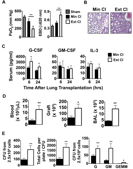
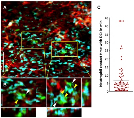
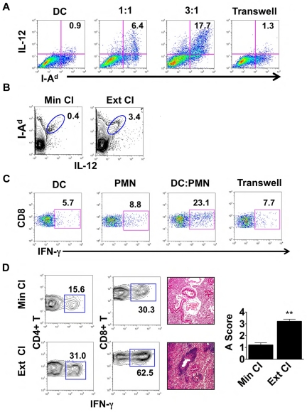
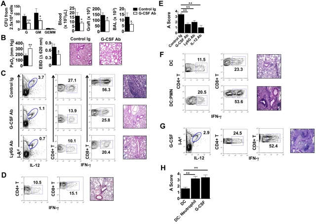
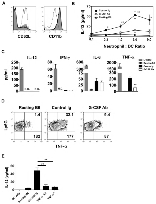
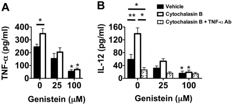
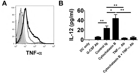
The mechanisms by which innate immune signals regulate alloimmune responses remain poorly understood. In the present study, we show by intravital 2-photon microscopy direct interactions between graft-infiltrating neutrophils and donor CD11c(+) dendritic cells (DCs) within orthotopic lung allografts immediately after reperfusion. Neutrophils isolated from the airways of lung transplantation recipients stimulate donor DCs in a contact-dependent fashion to augment their production of IL-12 and expand alloantigen-specific IFN-γ(+) T cells. DC IL-12 expression is largely regulated by degranulation and induced by TNF-α associated with the neutrophil plasma membrane. Extended cold ischemic graft storage enhances G-CSF-mediated granulopoiesis and neutrophil graft infiltration, resulting in exacerbation of ischemia-reperfusion injury after lung transplantation. Ischemia reperfusion injury prevents immunosuppression-mediated acceptance of mouse lung allografts unless G-CSF-mediated granulopoiesis is inhibited. Our findings identify granulopoiesis-mediated augmentation of alloimmunity as a novel link between innate and adaptive immune responses after organ transplantation.


Figures

Figure 1
Figure 1
Emergency granulopoiesis. (A) PaO2 (left) and Evans Blue Dye (EBD; right) exclusion in sham-operated B6 mice or 24 hours after Min CI or Ext CI Balb/c → B6 transplantation (N ≥ 5). (B) Histology from Min CI (n = 4) or Ext CI (n = 5) Balb/c → B6 lungs on POD 1. (C) Serum concentrations of indicated cytokines from Min CI or Ext CI Balb/c → B6 transplantations 6 and 24 hours after engraftment (n ≥ 4). (D) Neutrophil numbers in the blood, graft tissue, and BAL from Min CI or Ext CI Balb/c → B6 transplantations on POD 1 (n = 4). (E) BM cells were isolated from Min CI or Ext CI Balb/c → B6 lung transplantations 36 hours after engraftment and 2.5 × 10⁴ cells per plate were assayed in methylcellulose for CFU (left), total number of cells per plate to CFU (center), and CFU (right) differentiated as arising from granulocyte (G), granulocyte-macrophage (GM), and granulocyte-erythroid-macrophage-megakaryocyte (GEMM) progenitors. Data shown are from 1 representative experiment from 4 independently conducted experiments. Data for panels A through E represent the mean ± SEM. *P < .05; **P < .01.
Figure 2
Figure 2
Neutrophils make prolonged associations with donor-derived graft-resident. (A) Intravital 2P imaging of the distribution of donor DCs (green) and recipient neutrophils (blue) within lung grafts 1 hour after B10.BR CD11c-EYFP → B6 LysM-GFP transplantation. Blood vessels appear red (nontargeted 655-nm quantum dots). Scale bar indicates 60 μm. (B) Zoomed views show several neutrophils (yellow) making contact with CD11c+ DCs (white). Scale bar indicates 15 μm. (C) Measurement of neutrophil contact time with DCs over a 43.5-minute period approximately 1 hour after engraftment. The bar represents the mean neutrophil-DC contact time of 6.8 minutes. Some cells remained associated for the entire imaging period.
Figure 3
Figure 3
Neutrophils from lung recipients activate DCs in a contact-dependent manner. (A) IL-12 and MHC II (I-Ad) expression in Balb/c BMDCs cultured for 24 hours alone or with BAL neutrophils from Ext CI B6 → B6 lung transplantations at a BAL neutrophil to BMDC ratio of 1:1 or 3:1, or separated by a Transwell at a ratio of 3:1. (B) Percent abundance of intragraft IL-12+ I-Ad+ cells in Min CI or Ext CI Balb/c → B6 transplantations on POD 1. (C) IFN-γ expression after stimulation with Balb/c splenocytes in CD8+ T cells isolated from B6 mice 1 week after injection of immature Balb/c BMDCs (DC), BAL neutrophils (PMN) from Ext CI B6 → B6 transplantations, Balb/c BMDCs cultured with PMN, or Balb/c BMDCs cultured with PMN separated by a Transwell. (D) Percent abundance of intragraft IFN-γ+CD4+ and IFN-γ+CD8+ T cells (n ≥ 6; left), histology (n ≥ 6; center), and rejection scores (n = 5; right) in costimulation blockade-treated Min CI or Ext CI Balb/c → B6 transplantations on POD 7. Results are shown as means ± SEM. *P < .05; **P < .01. Data for panels A-E are representative of ≥ 3 independent experiments unless otherwise noted.
Figure 4
Figure 4
G-CSF blockade promotes the acceptance of lung allografts exposed to extended cold ischemia. (A) BM cells isolated from Ext CI Balb/c → B6 lung transplantations treated with control Ig or anti–G-CSF Ab 36 hours after engraftment and 2.5 × 10⁴ cells per plate were methylcellulose assayed for CFU-G, CFU-GM and CFU-GEMM numbers (n = 4; left). Neutrophil numbers in peripheral blood, lung tissue, and BAL from control Ig or anti–G-CSF Ab–treated Ext CI Balb/c → B6 transplantations 24 hours after engraftment (n = 4; right). (B) Analysis of PaO2 (n = 5; left), exclusion of EBD (center); and histology (n = 5; right) after engraftment. (C) Percent abundance of intragraft IL-12+ I-Ad+ cells on POD 1 (n = 3; left), percent abundance of intragraft IFN-γ+CD4+ and IFN-γ+CD8+ T cells on POD 7 (n ≥ 4; center) and representative histology (n ≥ 4; right) on POD 7 from control Ig–treated, G-CSF Ab–treated, or Ly6G Ab–treated Ext CI Balb/c → B6 transplantations that received costimulatory blockade. (D) Ext CI Balb/c → B6 transplantations that received costimulatory blockade and IL-12–neutralizing Abs (n = 4) and assessed on POD 7 for the percentage abundance of intragraft IFN-γ+ CD4+ and IFN-γ+ CD8+ T cells (n = 4; left) and representative histology (n = 4; right) (E) Rejection scoring of lung grafts on POD 7 from costimulatory blockade-treated Ext CI Balb/c → B6 recipients that received control Ig, G-CSF, Ly6G, or IL-12 Abs. (F) Percent abundance of intragraft IFN-γ+CD4+ and IFN-γ+CD8+ T cells (n ≥ 3; left) and representative histology (n ≥ 3; right) on POD 7 from costimulatory blockade-treated Min CI Balb/c → B6 transplantations that received an IV injection of either 5 × 10⁵ Balb/c BMDCs (DC) or 5 × 10⁵ Balb/c BMDCs activated by coculture with BAL neutrophils from Ext CI B6 → B6 transplantations (DC: Neutrophil). (G) Percent abundance of intragraft IL-12+I-Ad+ cells on POD 1 (n = 3; left), percent abundance of intragraft IFN-γ+CD4+ and IFN-γ+CD8+ T cells (n = 4; center) on POD 7 and histology on POD 7 (n = 4; right) from costimulatory blockade–treated Min CI Balb/c → B6 transplantations that received mouse recombinant G-CSF (10 μg) immediately after reperfusion. (H) Rejection scoring on POD 7 for costimulatory blockade–treated Min CI Balb/c → B6 transplantations that received DCs, DC:Neutrophil, or G-CSF. Data for panels A, B, E, and H represent the means ± SEM. *P < .05; **P < .01.
Figure 5
Figure 5
TNF-α from lung recipient neutrophils drives IL-12 expression in DCs. (A) Representative histogram (n = 4) of CD62L and CD11b expression of either neutrophils isolated from the airway of control Ig–treated (thick line) and G-CSF Ab-treated (solid line) Ext CI Balb/c → B6 lung recipients on POD 1 or neutrophils from resting B6 mouse BM (thin line). (B) Airway neutrophils from control Ig–treated or G-CSF Ab–treated Ext CI Balb/c → B6 lung recipients on POD1 or neutrophils from resting B6 mouse BM were cocultured with BMDCs in the indicated neutrophil to DC ratios for 24 hours and then supernatants were assessed for the accumulation of IL-12. Results are representative of 2 independently performed experiments. (C) BMDCs were stimulated with 10 ng/mL of LPS for 24 hours (LPS DC) or POD 1 airway neutrophils from control Ig–treated or G-CSF Ab–treated Ext CI Balb/c → B6 lung recipients or neutrophils from resting B6 mouse BM were cultured for 24 hours and then supernatants were analyzed for IL-12, IFN-γ, IL-6, and TNF-α. Results are representative of 3 independently performed experiments. (D) Intracellular TNF-α expression from resting B6 mouse BM neutrophils or airway neutrophils from control Ig–treated or G-CSF Ab–treated Ext CI Balb/c → B6 lung recipients on POD 1. Representative results from 4 independent experiments are shown, in which the upper right number indicates the percentage abundance of TNF-α+ cells and the lower right number is the mean fluorescence intensity of TNF-α expression. (E) IL-12 accumulation in the supernatants of BMDC culture or BMDC coculture with resting B6 neutrophils, POD 1 airway neutrophils from Ext CI Balb/c → B6 lung recipients, treated with control Ig or TNF-α Abs, or POD 1 airway neutrophils from Ext CI Balb/c → B6 TNF-α−/− lung recipients. Data are representative of 2 independent experiments. Data for panels B and E represent the means ± SEM. *P < .05; **P < .01.
Figure 6
Figure 6
Neutrophil degranulation increases TNF-α–mediated IL-12 expression in DCs. (A) Supernatant TNF-α accumulation of vehicle-treated, cytochalasin B-treated, genistein-treated, or genistein + cytochalasin B–treated POD1 airway neutrophils from Ext CI Balb/c → B6 lung recipients. Results are representative of 3 independent experiments. (B) Supernatant IL-12 accumulation in BMDC cocultures with vehicle-treated, cytochalasin B-treated, genistein-treated, or genistein + cytochalasin B–treated POD1 airway neutrophils from Ext CI Balb/c → B6 lung recipients cultured in the absence or presence of TNF-α Abs. Results are representative of 3 independent experiments. Data for panels A and B represent the mean ± SEM. *P < .05; **P < .01.
Figure 7
Figure 7
TNF-α is associated with the neutrophil plasma membranes and stimulates IL-12 expression in DCs. (A) Histogram of surface TNF-α expression on POD 1 airway neutrophils from unstimulated (thick gray line) or cytochalasin B–stimulated (thick black line) control Ig–treated Ext CI Balb/c → B6 lung recipients or (thin black line) G-CSF Ab–treated Ext CI Balb/c → B6 lung recipients or (shaded) Ext CI Balb/c → B6 TNF-α−/− lung recipients. Results are representative of 2 independent experiments. (B) BMDC culture (DCs only) or BMDCs cocultured with unstimulated (control Ig) or cytochalasin B–stimulated (cytochalasin B) POD 1 Ext CI Balb/c → B6 lung recipient airway neutrophil plasma membranes in the absence or presence of TNF-α Ab (TNF-α Ab or cytochalasin B + TNF-α Ab, respectively) or with POD 1 G-CSF Ab–treated Ext CI Balb/c → B6 lung recipient airway neutrophil plasma membranes (G-CSF Ab). One day later, IL-12 accumulation was assessed in culture supernatants. Results are representative of 2 independent experiments. Data represent the means ± SEM. *P < .05; **P < .01.

References

    1. Christie JD, Kotloff RM, Ahya VN, et al. The effect of primary graft dysfunction on survival after lung transplantation. Am J Respir Crit Care Med. 2005;171(11):1312–1316. - PMC - PubMed
    1. Kreisel DKA, Puri V, Guthrie TJ, Trulock EP, Meyers BF, Patterson GA. Short- and long-term outcomes of 1000 adult lung transplant recipients at a single center. J Thorac Cardiovasc Surg. 2011;141(1):215–222. - PubMed
    1. de Perrot M, Liu M, Waddell TK, Keshavjee S. Ischemia-reperfusion-induced lung injury. Am J Respir Crit Care Med. 2003;167(4):490–511. - PubMed
    1. Kreisel D, Sugimoto S, Tietjens J, et al. Bcl3 prevents acute inflammatory lung injury in mice by restraining emergency granulopoiesis. J Clin Invest. 2011;121(1):265–276. - PMC - PubMed
    1. Hirai H, Zhang P, Dayaram T, et al. C/EBPbeta is required for ‘emergency’ granulopoiesis. Nat Immunol. 2006;7(7):732–739. - PubMed

Publication types

MeSH terms

Substances