. 2011 Nov 17;118(20):5498-505.
doi: 10.1182/blood-2011-07-365825. Epub 2011 Oct 4.

Positive feedback between PGE2 and COX2 redirects the differentiation of human dendritic cells toward stable myeloid-derived suppressor cells

Affiliations

Positive feedback between PGE2 and COX2 redirects the differentiation of human dendritic cells toward stable myeloid-derived suppressor cells

Natasa Obermajer et al. Blood. .

Abstract

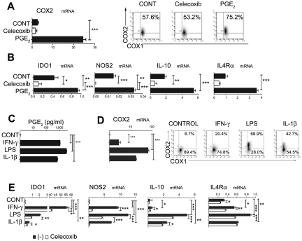
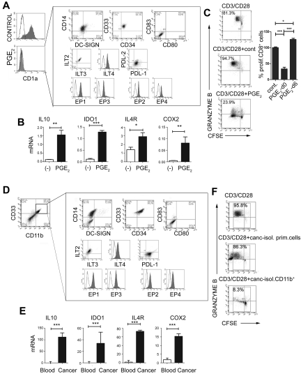
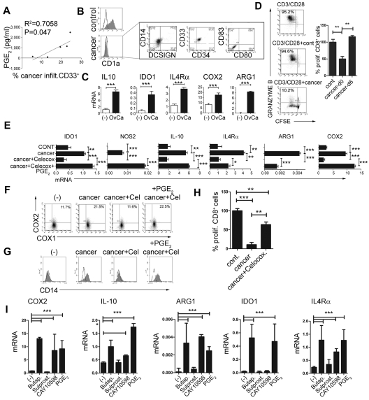
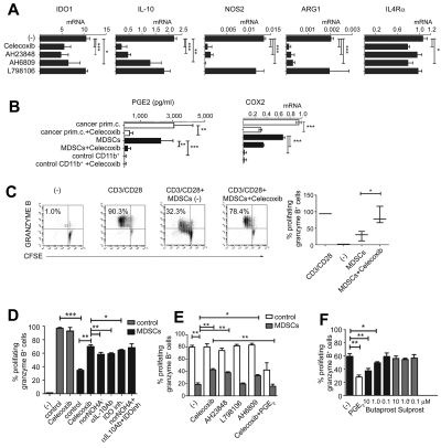
Dendritic cells (DCs) and myeloid-derived suppressor cells (MDSCs) show opposing roles in the immune system. In the present study, we report that the establishment of a positive feedback loop between prostaglandin E(2) (PGE(2)) and cyclooxygenase 2 (COX2), the key regulator of PGE(2) synthesis, represents the determining factor in redirecting the development of CD1a(+) DCs to CD14(+)CD33(+)CD34(+) monocytic MDSCs. Exogenous PGE(2) and such diverse COX2 activators as lipopolysaccharide, IL-1β, and IFNγ all induce monocyte expression of COX2, blocking their differentiation into CD1a(+) DCs and inducing endogenous PGE(2), IDO1, IL-4Rα, NOS2, and IL-10, typical MDSC-associated suppressive factors. The addition of PGE(2) to GM-CSF/IL-4-supplemented monocyte cultures is sufficient to induce the MDSC phenotype and cytotoxic T lymphocyte (CTL)-suppressive function. In accordance with the key role of PGE(2) in the physiologic induction of human MDSCs, the frequencies of CD11b(+)CD33(+) MDSCs in ovarian cancer are closely correlated with local PGE(2) production, whereas the cancer-promoted induction of MDSCs is strictly COX2 dependent. The disruption of COX2-PGE(2) feedback using COX2 inhibitors or EP2 and EP4 antagonists suppresses the production of MDSC-associated suppressive factors and the CTL-inhibitory function of fully developed MDSCs from cancer patients. The central role of COX2-PGE(2) feedback in the induction and persistence of MDSCs highlights the potential for its manipulation to enhance or suppress immune responses in cancer, autoimmunity, or transplantation.


Figures

Figure 1
Figure 1
Inhibition of DC differentiation by PGE2, proinflammatory cytokines, and TLR ligands is associated with the induction of endogenous COX2- and MDSC-associated suppressive factors. (A) Expression of COX2 mRNA (panel A left) and protein (panel A right) levels is induced by synthetic PGE2, initiating a positive feedback loop in iMCs. Regulation of COX1 and COX2 expression by synthetic PGE2 was analyzed after 6-10 hours. (B) Induction of immunosuppressive factors IL-10, NOS2, IDO1, and IL-4Rα by synthetic PGE2. (C-D) Induction of PGE2 secretion (C) and COX2 mRNA (D left) and protein (D right) by the COX2 activators LPS (TLR4 ligand), IL-1β, and IFN-γ. (E) Celecoxib suppresses the induction of immunosuppressive factors (IDO1, IL-10, NOS2, and IL-4Rα) by COX2 activators. All data (panels A-E) were confirmed in at least 3 independent experiments. Histograms present data from a single representative experiment with different donors as mean ± SD. *P < .05; **P < .01; and ***P < .001.
Figure 2
Figure 2
PGE2 redirects DC differentiation and induces CD14+CD33+CD34+ cells with the phenotype and function of monocytic MDSCs isolated from cancer patients. (A) Phenotype of PGE2-induced CD1aCD14+CD80CD83 MDSCs expressing inhibitory molecules ILT2, ILT3, ILT4, PDL-1, but not PDL-2. PGE2-induced MDSCs express E-prostanoid receptors (labeled with α-EP1–, α-EP3–, sec.Alexa488, α-EP2–, and α-EP4–PE). (B) Expression of immunosuppressive factors IL-10, IDO1, IL-4Rα, and COX2 in PGE2-induced MDSCs (see supplemental Figure 1A for corresponding protein levels of IDO and IL-10). (C) Immunosuppressive effects of PGE2-induced MDSCs on allogeneic naive CFSE-labeled CD8+ T cells primed by CD3/CD28 and stained for granzyme B. Left panel: Percentages indicate the fraction of proliferating granzyme B+ (marker of CTL status) CD8+ cells. Right panel: Percentage of proliferating CD8+ T cells in the presence of PGE2-induced MDSCs (PGE2-d0) and PGE2-conditioned DCs (PGE2-d6). (D-F) MDSC phenotype and function of CD11b+ cells isolated from cancer ascites. (D) Characterization of cells from cancer ascites either before (left panel) or after (right panel box) isolation of CD11b+ cells. Note the high percentage of CD11b+ cells (8.9%-50.0%, mean 24.2%, n = 7) within the cancer-infiltrating primary cell population. (E) mRNA levels of IL-10, IDO1, IL-4Rα, and COX2 in CD11b+ cells isolated from cancer (see supplemental Figure 1B for corresponding protein levels of IDO and COX2) compared with CD11b+ cells isolated from blood (ascites-isolated, n = 7; control blood-isolated, n = 5). (F) Suppression of CFSE-labeled allogeneic naive CD8+ T-cell proliferation (CD3/CD28 stimulation) in the presence or absence of primary cells or CD11b+ cells isolated from cancer (ie, MDSCs isolated from cancer; n = 7). Percentages indicate the fraction of proliferating granzyme B+CD8+ cells. The gray squares represent the lymphocyte-specific gates used to exclude (CFSE-unlabeled) MDSCs. All data (panels A-F) were confirmed in at least 3 independent experiments. Histograms present data of a single representative experiment with different donors as means ± SD. *P < .05; **P < .01; and ***P < .001.
Figure 3
Figure 3
PGE2 mediates the enhanced development of MDSCs in human cancer environment. (A) Correlation between the PGE2 production and the frequencies of cancer-infiltrating CD11b+CD33+ cells from different patients. The percentage of cancer-infiltrating CD11b+CD33+ cells was determined by flow cytometry (n = 5 patients). The regression line and corresponding R2 value are shown. (B) MDSC phenotype induced in GM-CSF+IL-4–cultured monocytes by membrane-permeable soluble factor(s) produced by cancer-infiltrating cells. Similar data were obtained using the CM from cancer-infiltrating cells and in a Transwell system (see supplemental Figure 3 for experimental design). Left panel: Suppression of DC differentiation by CM from cancer-infiltrating cells (manifested by loss of the DC marker CD1a). Right panel: Induction of the CD1aCD14+DCSIGNCD80CD83 MDSC phenotype. (C) mRNA levels of IL-10, arginase 1 (ARG1), IDO1, IL-4Rα, and COX2 in cancer-induced MDSCs. (D) Suppressed CD3/CD28–induced proliferation of granzyme B+ CTL (percentages) in the presence of cancer-induced MDSCs. Left panel: Percentages indicate the fraction of proliferating granzyme B+CD8+ cells. Right panel: Percentage of proliferating CD8+ T cells in the presence of cancer-induced MDSCs (cancer d0) and cancer-conditioned DCs (cancer d6). (E) Induction of immunosuppressive factors by cancer-associated PGE2. (F) Induction of CD14+ MDSCs by CM from cancer-infiltrating cells is suppressed by COX2 inhibition and restored by synthetic PGE2. (G) Regulation of COX1 and COX2 expression by CM from cancer-infiltrating cells generated in the presence or absence of celecoxib and/or synthetic PGE2 and analyzed after 6-10 hours. (H) Immunosuppressive effects of cancer-induced MDSCs on naive CFSE-labeled CD8+ T cells primed by CD3/CD28 and stained for granzyme B. Cancer-infiltrating primary cell CM was generated in the presence or absence of the COX2 inhibitor celecoxib. (I) Induction of immunosuppressive factors by PGE2, the EP4 agonist CAY10598, the EP2 agonist butaprost, but not the EP3/1 agonist sulprostone. All data (panels A-I) were confirmed in 3-7 independent experiments. Histograms present data from a single representative experiment with different donors as mean ± SD.
Figure 4
Figure 4
Functional stability of MDSCs isolated from cancer patients requires continuous PGE2-COX2 feedback involving EP2 and EP4 signaling. (A) Expression of immunosuppressive factors in cancer-isolated CD11b+ cells pretreated (24 hours) or not with celecoxib, the EP4 antagonist AH23848, the EP2/1 antagonist AH6809, and the EP3 antagonist L798106. (B) PGE2 production and COX2 expression in primary cells (white bars) and CD11b+ cells (black bars) isolated from cancer, compared with control CD11b+ cells isolated from blood treated or not with celecoxib. Measurements were performed in triplicates. (C) Celecoxib pretreatment of MDSCs isolated from cancer abolishes their suppressive impact on CD3/CD28–activated naive CD8+ T cells. (D) Inhibition of COX2, arginase-1, IL-10, or IDO1 counteracts the suppressive functions of MDSCs isolated from cancer (n = 3). (E) Celecoxib, the EP4 antagonist AH23848, and the EP2/1 antagonist AH6809, but not the EP3 antagonist L798106, similarly reverse the suppressive functions of MDSCs. The addition of synthetic PGE2 to celecoxib-pretreated MDSCs isolated from cancer restores immunosuppressive functions (n = 3). Neither celecoxib nor the EP antagonists showed any cytotoxic effects at the concentrations used. (F) CD3/CD28 activation of CD8+ T cells (absence of MDSCs) ± 1μM PGE2 or different concentrations of the commercially available agonists butaprost (EP2) or sulprostone (EP3/1 agonist, negative control). All data (panels A-F) were confirmed in at least 3 independent experiments. Histograms present data from a single representative experiment with different donors as means ± SD.

References

    1. Pulendran B, Palucka K, Banchereau J. Sensing pathogens and tuning immune responses. Science. 2001;293(5528):253–256. - PubMed
    1. Palucka K, Ueno H, Roberts L, Fay J, Banchereau J. Dendritic cells: are they clinically relevant? Cancer J. 2010;16(4):318–324. - PMC - PubMed
    1. Torres-Aguilar H, Blank M, Jara LJ, Shoenfeld Y. Tolerogenic dendritic cells in autoimmune diseases: crucial players in induction and prevention of autoimmunity. Autoimmun Rev. 2010;10(1):8–17. - PubMed
    1. Pulendran B, Tang H, Manicassamy S. Programming dendritic cells to induce T(H)2 and tolerogenic responses. Nat Immunol. 2010;11(8):647–655. - PubMed
    1. Condamine T, Gabrilovich DI. Molecular mechanisms regulating myeloid-derived suppressor cell differentiation and function. Trends Immunol. 2011;32(1):19–25. - PMC - PubMed

Publication types

MeSH terms