

MRI analysis in temporal lobe epilepsy: cortical thinning and white matter disruptions are related to side of seizure onset
- PMID: 21972957
- PMCID: PMC3230670
- DOI: 10.1111/j.1528-1167.2011.03278.x
MRI analysis in temporal lobe epilepsy: cortical thinning and white matter disruptions are related to side of seizure onset
Abstract
Purpose: Past studies reported more widespread structural brain abnormalities in patients with left compared to right temporal lobe epilepsy (TLE), but the profile of these differences remains unknown. This study investigated the relationship between cortical thinning, white matter compromise, epilepsy variables, and the side of seizure onset, in patients with TLE.
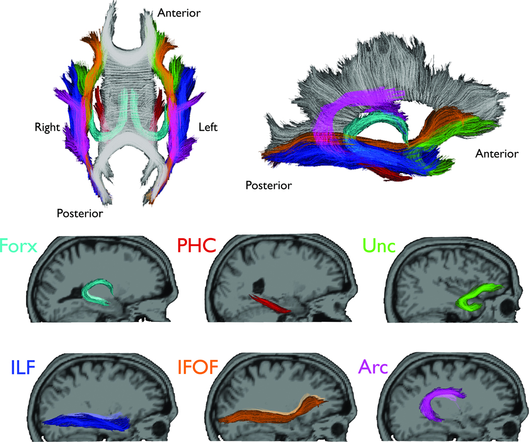
Methods: We performed diffusion tensor imaging tractography and cortical thickness analyses of 18 patients with left TLE (LTLE), 18 patients with right TLE (RTLE), and 36 controls. We investigated the relationship among brain structural abnormalities, side of seizure onset, age of seizure onset, and disease duration.
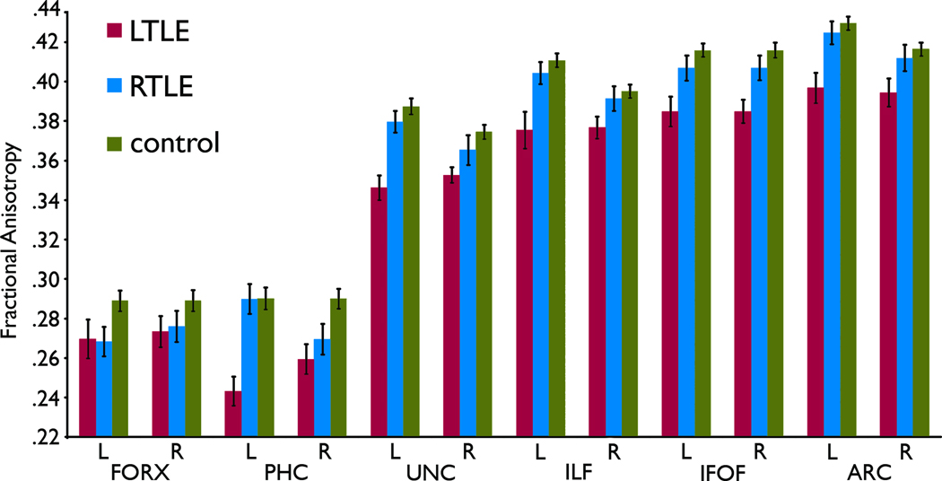
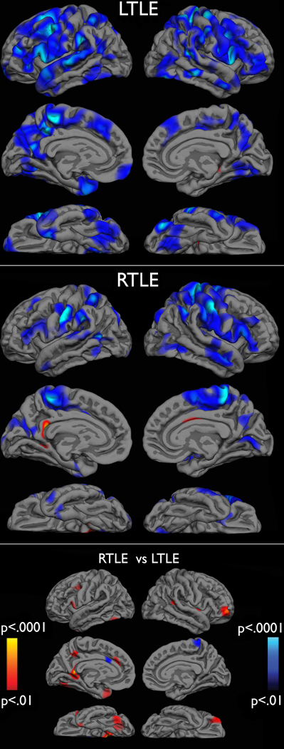
Key findings: Patients with TLE displayed cortical thinning and white matter compromise, predominately on the side ipsilateral to the seizure onset. Relative to RTLE, patients with LTLE showed more widespread abnormalities, particularly in white matter fiber tracts. Greater compromise in white matter integrity was associated with earlier age of seizure onset, whereas cortical thinning was marginally associated with disease duration.
Significance: These data support previous findings of LTLE showing greater structural compromise than RTLE, and suggest that mechanisms may not be uniform for gray and white matter compromise in patients with LTLE and RTLE. These results may indicate that LTLE is different from RTLE, possibly due to greater vulnerability of the left hemisphere to early injury and the progressive effects of seizures.
Wiley Periodicals, Inc. © 2011 International League Against Epilepsy.
Figures
References
-
- Bell BD, Davies KG. Anterior temporal lobectomy, hippocampal sclerosis, and memory: recent neuropsychological findings. Neuropsychol Rev. 1998;8:25–41. - PubMed
-
- Bernasconi N, Duchesne S, Janke A, Lerch J, Collins DL, Bernasconi A. Whole-brain voxel-based statistical analysis of gray matter and white matter in temporal lobe epilepsy. Neuroimage. 2004;23:717–723. - PubMed
-
- Bernasconi N, Natsume J, Bernasconi A. Progression in temporal lobe epilepsy: differential atrophy in mesial temporal structures. Neurology. 2005;65:223–228. - PubMed
-
- Bernhardt BC, Bernasconi N, Concha L, Bernasconi A. Cortical thickness analysis in temporal lobe epilepsy: reproducibility and relation to outcome. Neurology. 2010;74:1776–1784. - PubMed
Publication types
MeSH terms
Grants and funding
LinkOut - more resources
Full Text Sources
Medical
Miscellaneous
