. 2011 Dec;278(24):4845-59.
doi: 10.1111/j.1742-4658.2011.08385.x. Epub 2011 Nov 3.

Identification and verification of heat shock protein 60 as a potential serum marker for colorectal cancer

Affiliations
Free PMC article

Identification and verification of heat shock protein 60 as a potential serum marker for colorectal cancer

Céline Hamelin et al. FEBS J. 2011 Dec.
Free PMC article

Abstract

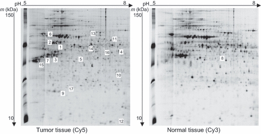
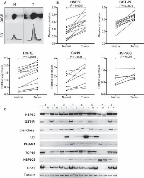
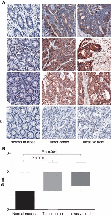
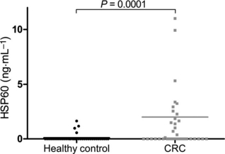
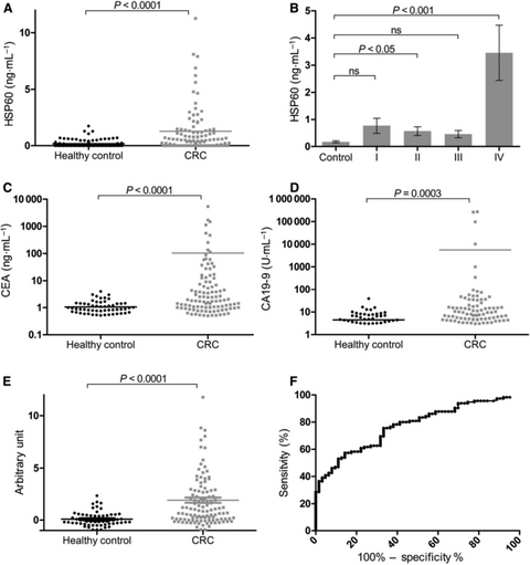
Colorectal cancer (CRC) is a major public health issue worldwide, and novel tumor markers may contribute to its efficient management by helping in early detection, prognosis or surveillance of disease. The aim of our study was to identify new serum biomarkers for CRC, and we followed a phased biomarker discovery and validation process to obtain an accurate preliminary assessment of potential clinical utility. We compared colonic tumors and matched normal tissue from 15 CRC patients, using two-dimensional difference gel electrophoresis (2D-DIGE), and identified 17 proteins that had significant differential expression. These results were further confirmed by western blotting for heat shock protein (HSP) 60, glutathione-S-transferase Pi, α-enolase, T-complex protein 1 subunit β, and leukocyte elastase inhibitor, and by immunohistochemistry for HSP60. Using mAbs raised against HSP60, we developed a reliable (precision of 5-15%) and sensitive (0.3 ng·mL(-1)) immunoassay for the detection of HSP60 in serum. Elevated levels of HSP60 were found in serum from CRC patients in two independent cohorts; the receiver-operating characteristic curve obtained in 112 patients with CRC and 90 healthy controls had an area under the curve (AUC) of 0.70, which was identical to the AUC of carcinoembryonic antigen. Combination of serum markers improved clinical performance: the AUC of a three-marker logistic regression model combining HSP60, carcinoembryonic antigen and carbohydrate antigen 19-9 reached 0.77. Serum HSP60 appeared to be more specific for late-stage CRC; therefore, future studies should evaluate its utility for determining prognosis or monitoring therapy rather than early detection.


Figures

Fig. 1
Fig. 1
Representative 2D-DIGE maps of colonic tissue (patient 6). Soluble proteins extracted from colon tumor (Cy5) and matched normal tissue (Cy3) were labeled with the indicated dyes, mixed with Cy2-labeled internal standard, and subjected to IEF on pH 5-8 IPG strips. Protein samples were then separated on large-format 7.7–16.5% gradient SDS/PAGE gels. Molecular mass separation is 150–10 kDa (top to bottom). Numbered spots indicate proteins that have statistically significant differential expression between tumor tissue and adjacent normal mucosa (fold-change over 1.5 and P < 0.05 with Wilcoxon signed-rank test). MALDI-TOF MS identification results for these spots are shown in Table 2.
Fig. 2
Fig. 2
Western blot qualification of differentially expressed protein spots. (A) 2D-DIGE image and corresponding 3D simulation of the HSP60 spot in a matched tissue sample. N, normal tissue; T, tumoral tissue. (B) Relative expression of HSP60, GST-Pi, TCP1β, CK19 and HSP90β in paired CRC samples analyzed by 2D-DIGE. Relative expression corresponds to the spot volume determined with imagemaster 2d-platinium software, transformed into logarithm base 2, and normalized with the corresponding spot volume of the internal standard image (Cy2). Comparisons were performed with the Wilcoxon signed-rank test. (C) Western blot analysis of protein expression in eight independent tissue sample pairs. Tubulin was used as loading control.
Fig. 3
Fig. 3
(A) Representative immunohistochemical staining images of HSP60 in normal colonic mucosa, tumor center, and tumor just behind the invasive front; magnification, × 20. For negative controls (bottom panels, Ctr), primary antibody against HSP60 was replaced by an irrelevant mouse IgG. (B) Comparison of HSP60 staining scores between matched normal mucosa, invasive front and tumor center in a series of 20 specimens from CRC patients. Analysis of variance with Friedman's test showed significant differences in the dataset (P < 0.0001). Pairwise post hoc comparisons were performed with Dunn's multiple comparison test, and the corresponding P-values are shown.
Fig. 4
Fig. 4
Serum levels of HSP60 in the qualification cohort, 40 healthy controls and 40 CRC patients, measured by ELISA. Serum HSP60 levels were significantly elevated in CRC patients (P = 0.0001, one-tailed Mann–Whitney test). The gray line represents the mean HSP60 concentration for the CRC group.
Fig. 5
Fig. 5
Serum levels of HSP60 and other CRC markers in the verification cohort. (A) HSP60, n = 202, AUC = 0.70. (C) CEA, n = 175, AUC = 0.70. (D) CA19-9, n = 175, AUC = 0.65. (E) Three-marker combination calculated with a logistic regression model, expressed in arbitrary units, n = 175. Mean marker concentrations are represented by lines. Control and cancer groups were compared by use of the one-tailed Mann–Whitney test. (F) Receiver-operating characteristic curve of the three-marker combination, AUC = 0.77. (B) HSP60 concentration according to CRC stage (I–IV). Data are means ± standard errors. Analysis of variance with Friedman's test indicated significant differences in HSP60 levels between groups (P < 0.0001). Pairwise comparisons were performed with Dunn's multiple comparison test, and the corresponding P-values are shown.

Similar articles

Cited by

References

    1. Jemal A, Bray F, Center MM, Ferlay J, Ward E, Forman D. Global cancer statistics. CA Cancer J Clin. 2011;61:69–90. - PubMed
    1. Walsh JM, Terdiman JP. Colorectal cancer screening: clinical applications. JAMA. 2003;289:1297–1302. - PubMed
    1. Burt RW. Colorectal cancer screening. Curr Opin Gastroenterol. 2010;26:466–470. - PubMed
    1. Lieberman DA, Weiss DG, Bond JH, Ahnen DJ, Garewal H, Chejfec G. Use of colonoscopy to screen asymptomatic adults for colorectal cancer. Veterans Affairs Cooperative Study Group 380. N Engl J Med. 2000;343:162–168. - PubMed
    1. Nelson DB, McQuaid KR, Bond JH, Lieberman DA, Weiss DG, Johnston TK. Procedural success and complications of large-scale screening colonoscopy. Gastrointest Endosc. 2002;55:307–314. - PubMed