

Pattern of forebrain activation in high novelty-seeking rats following aggressive encounter
- PMID: 21974861
- PMCID: PMC3200440
- DOI: 10.1016/j.brainres.2011.08.033
Pattern of forebrain activation in high novelty-seeking rats following aggressive encounter
Abstract
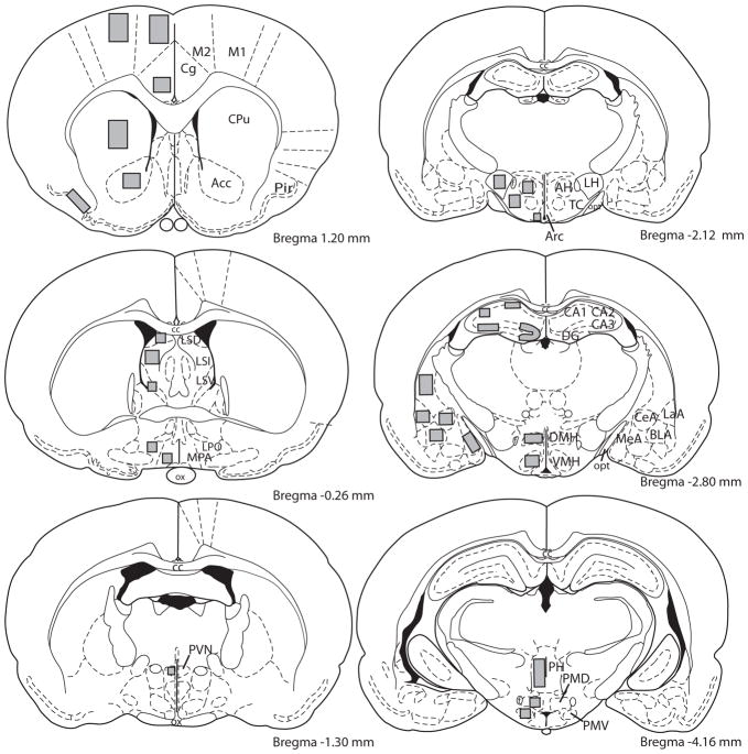
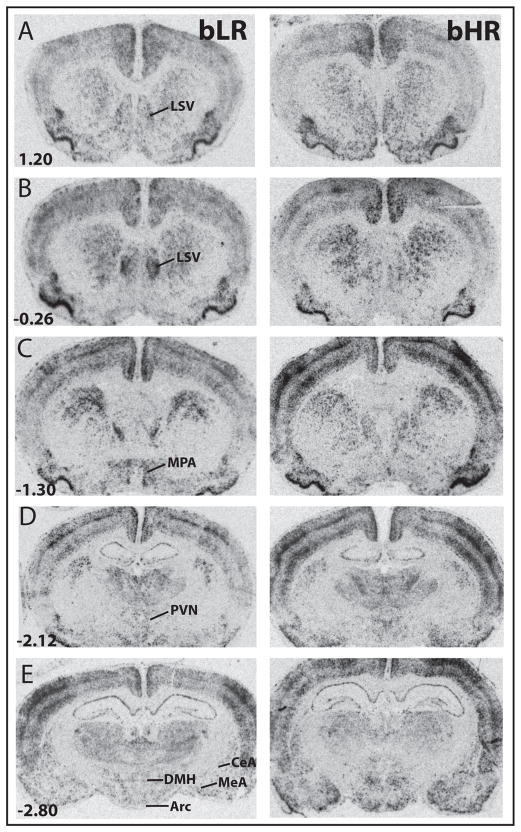
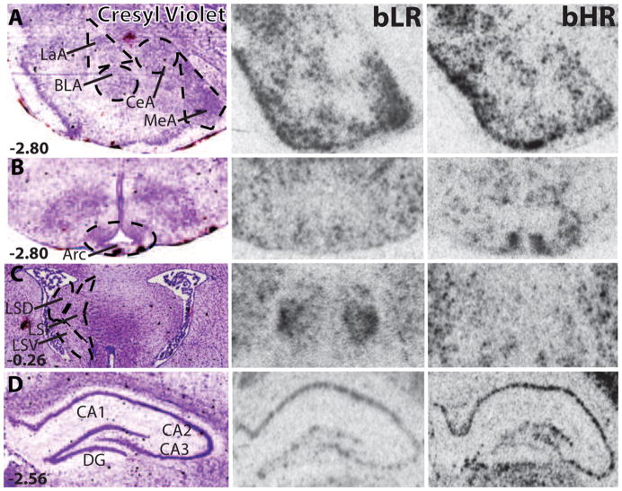
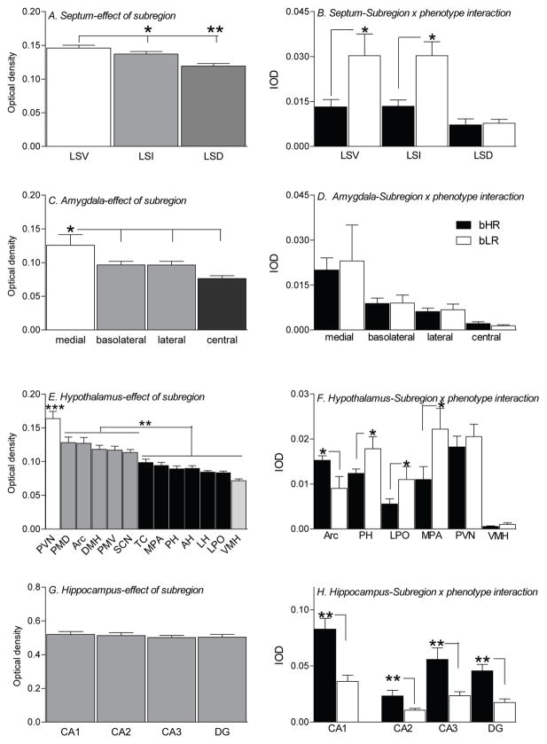
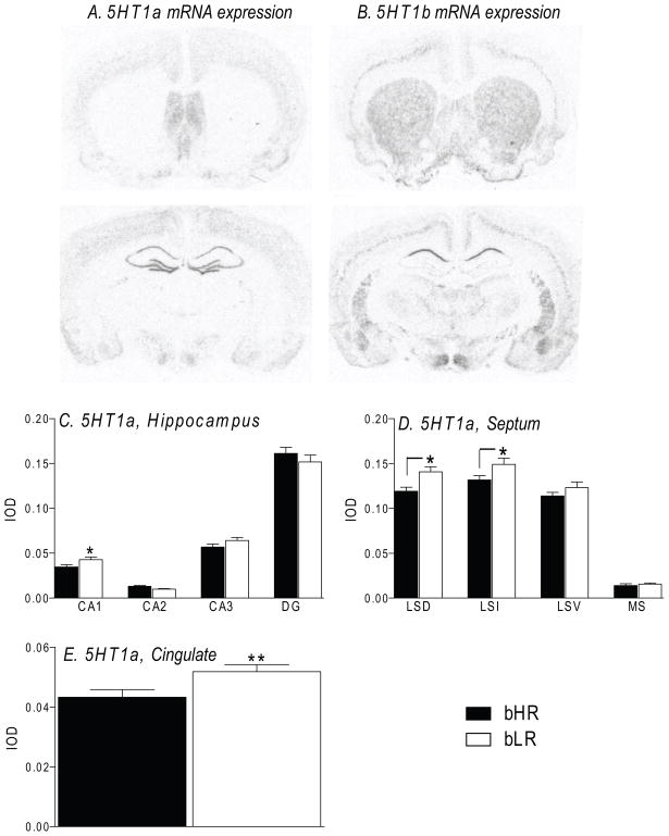
We have previously demonstrated that selectively-bred High (bHR) and Low (bLR) novelty-seeking rats exhibit agonistic differences, with bHRs acting in a highly aggressive manner when facing homecage intrusion. In order to discover the specific neuronal pathways responsible for bHRs' high levels of aggression, the present study compared c-fos mRNA expression in several forebrain regions of bHR/bLR males following this experience. bHR/bLR males were housed with female rats for 2 weeks, and then the females were replaced with a male intruder for 10 min. bHR/bLR residents were subsequently sacrificed by rapid decapitation, and their brains were removed and processed for c-fos in situ hybridization. Intrusion elicited robust c-fos mRNA expression in both phenotypes throughout the forebrain, including the septum, amygdala, hippocampus, cingulate cortex, and the hypothalamus. However, bHRs and bLRs exhibited distinct activation patterns in select areas. Compared to bHR rats, bLRs expressed greater c-fos in the lateral septum and within multiple hypothalamic nuclei, while bHRs showed greater activation in the arcuate hypothalamic nucleus and in the hippocampus. No bHR/bLR differences in c-fos expression were detected in the amygdala, cortical regions, and striatum. We also found divergent 5-HT1A receptor mRNA expression within some of these same areas, with bLRs having greater 5-HT1A, but not 5-HT1B, receptor mRNA levels in the septum, hippocampus and cingulate cortex. These findings, together with our earlier work, suggest that bHRs exhibit altered serotonergic functioning within select circuits during an aggressive encounter.
Copyright © 2011 Elsevier B.V. All rights reserved.
Conflict of interest statement
There are no biomedical financial interests or conflicts of interest for any of the authors.
Figures
Similar articles
-
High novelty-seeking predicts aggression and gene expression differences within defined serotonergic cell groups.Brain Res. 2011 Oct 24;1419:34-45. doi: 10.1016/j.brainres.2011.08.038. Epub 2011 Aug 22. Brain Res. 2011. PMID: 21925645 Free PMC article.
-
Inborn differences in environmental reactivity predict divergent diurnal behavioral, endocrine, and gene expression rhythms.Psychoneuroendocrinology. 2012 Feb;37(2):256-69. doi: 10.1016/j.psyneuen.2011.06.010. Epub 2011 Jul 20. Psychoneuroendocrinology. 2012. PMID: 21775066 Free PMC article.
-
Developmental underpinnings of differences in rodent novelty-seeking and emotional reactivity.Eur J Neurosci. 2011 Sep;34(6):994-1005. doi: 10.1111/j.1460-9568.2011.07811.x. Epub 2011 Aug 22. Eur J Neurosci. 2011. PMID: 21864320 Free PMC article.
-
Resilience to Stress: Lessons from Rodents about Nature versus Nurture.Neuroscientist. 2022 Jun;28(3):283-298. doi: 10.1177/1073858421989357. Epub 2021 Feb 10. Neuroscientist. 2022. PMID: 33567987 Free PMC article. Review.
-
Antecedents and consequences of drug abuse in rats selectively bred for high and low response to novelty.Neuropharmacology. 2014 Jan;76 Pt B(0 0):425-36. doi: 10.1016/j.neuropharm.2013.04.033. Epub 2013 Apr 29. Neuropharmacology. 2014. PMID: 23639434 Free PMC article. Review.
Cited by
-
Differences in microglia morphological profiles reflect divergent emotional temperaments: insights from a selective breeding model.Transl Psychiatry. 2022 Mar 15;12(1):105. doi: 10.1038/s41398-022-01821-4. Transl Psychiatry. 2022. PMID: 35292624 Free PMC article.
-
Early-life exposure to the SSRI paroxetine exacerbates depression-like behavior in anxiety/depression-prone rats.Neuroscience. 2015 Jan 22;284:775-797. doi: 10.1016/j.neuroscience.2014.10.044. Epub 2014 Nov 4. Neuroscience. 2015. PMID: 25451292 Free PMC article.
-
Adolescent environmental enrichment induces social resilience and alters neural gene expression in a selectively bred rodent model with anxious phenotype.Neurobiol Stress. 2024 May 30;31:100651. doi: 10.1016/j.ynstr.2024.100651. eCollection 2024 Jul. Neurobiol Stress. 2024. PMID: 38933284 Free PMC article.
-
Independent effects of early-life experience and trait aggression on cardiovascular function.Am J Physiol Regul Integr Comp Physiol. 2016 Aug 1;311(2):R272-86. doi: 10.1152/ajpregu.00505.2015. Epub 2016 Jun 8. Am J Physiol Regul Integr Comp Physiol. 2016. PMID: 27280432 Free PMC article.
-
Coping Style of Pigs Is Associated With Different Behavioral, Neurobiological and Immune Responses to Stressful Challenges.Front Behav Neurosci. 2019 Aug 1;13:173. doi: 10.3389/fnbeh.2019.00173. eCollection 2019. Front Behav Neurosci. 2019. PMID: 31417378 Free PMC article.
References
-
- Adamec RE, Stark-Adamec C. Partial kindling and emotional bias in the cat: lasting aftereffects of partial kindling of the ventral hippocampus. I. Behavioral changes. Behav Neural Biol. 1983;38:205–22. - PubMed
-
- Adamec RE. Partial kindling of the ventral hippocampus: identification of changes in limbic physiology which accompany changes in feline aggression and defense. Physiology & Behavior. 1991;49:443–53. - PubMed
-
- Bandler R, Depaulis A, Vergnes M. Identification of midbrain neurones mediating defensive behaviour in the rat by microinjections of excitatory amino acids. Behav Brain Res. 1985;15:107–19. - PubMed
-
- Blanchard DC, Blanchard RJ, Lee EM, Nakamura S. Defensive behaviors in rats following septal and septal--amygdala lesions. J Comp Physiol Psychol. 1979;93:378–90. - PubMed
-
- Brown CS, Kent TA, Bryant SG, Gevedon RM, Campbell JL, Felthous AR, Barratt ES, Rose RM. Blood platelet uptake of serotonin in episodic aggression. Psychiatry Res. 1989;27:5–12. - PubMed
Publication types
MeSH terms
Grants and funding
LinkOut - more resources
Full Text Sources
