

Regional contrast agent quantification in a mouse model of myocardial infarction using 3D cardiac T1 mapping
- PMID: 21974927
- PMCID: PMC3207957
- DOI: 10.1186/1532-429X-13-56
Regional contrast agent quantification in a mouse model of myocardial infarction using 3D cardiac T1 mapping
Abstract
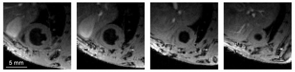
Background: Quantitative relaxation time measurements by cardiovascular magnetic resonance (CMR) are of paramount importance in contrast-enhanced studies of experimental myocardial infarction. First, compared to qualitative measurements based on signal intensity changes, they are less sensitive to specific parameter choices, thereby allowing for better comparison between different studies or during longitudinal studies. Secondly, T1 measurements may allow for quantification of local contrast agent concentrations. In this study, a recently developed 3D T1 mapping technique was applied in a mouse model of myocardial infarction to measure differences in myocardial T1 before and after injection of a liposomal contrast agent. This was then used to assess the concentration of accumulated contrast agent.
Materials and methods: Myocardial ischemia/reperfusion injury was induced in 8 mice by transient ligation of the LAD coronary artery. Baseline quantitative T1 maps were made at day 1 after surgery, followed by injection of a Gd-based liposomal contrast agent. Five mice served as control group, which followed the same protocol without initial surgery. Twenty-four hours post-injection, a second T1 measurement was performed. Local ΔR1 values were compared with regional wall thickening determined by functional cine CMR and correlated to ex vivo Gd concentrations determined by ICP-MS.
Results: Compared to control values, pre-contrast T1 of infarcted myocardium was slightly elevated, whereas T1 of remote myocardium did not significantly differ. Twenty-four hours post-contrast injection, high ΔR1 values were found in regions with low wall thickening values. However, compared to remote tissue (wall thickening > 45%), ΔR1 was only significantly higher in severe infarcted tissue (wall thickening < 15%). A substantial correlation (r = 0.81) was found between CMR-based ΔR1 values and Gd concentrations from ex vivo ICP-MS measurements. Furthermore, regression analysis revealed that the effective relaxivity of the liposomal contrast agent was only about half the value determined in vitro.
Conclusions: 3D cardiac T1 mapping by CMR can be used to monitor the accumulation of contrast agents in contrast-enhanced studies of murine myocardial infarction. The contrast agent relaxivity was decreased under in vivo conditions compared to in vitro measurements, which needs consideration when quantifying local contrast agent concentrations.
Figures


Similar articles
-
AUR Memorial Award. Identification of myocardial cell death in reperfused myocardial injury using dual mechanisms of contrast-enhanced magnetic resonance imaging.Acad Radiol. 1994 Dec;1(4):319-25. doi: 10.1016/s1076-6332(12)80001-8. Acad Radiol. 1994. PMID: 9419506
-
Quantitative T1 Mapping for Detecting Microvascular Obstruction in Reperfused Acute Myocardial Infarction: Comparison with Late Gadolinium Enhancement Imaging.Korean J Radiol. 2020 Aug;21(8):978-986. doi: 10.3348/kjr.2019.0736. Korean J Radiol. 2020. PMID: 32677382 Free PMC article.
-
T1-relaxation kinetics of extracellular, intracellular and intravascular MR contrast agents in normal and acutely reperfused infarcted myocardium using echo-planar MR imaging.Eur Radiol. 2000;10(2):310-8. doi: 10.1007/s003300050050. Eur Radiol. 2000. PMID: 10663763
-
The use of Gd-DTPA as a marker of myocardial viability in reperfused acute myocardial infarction.Int J Cardiovasc Imaging. 2001 Oct;17(5):395-404. doi: 10.1023/a:1011989626052. Int J Cardiovasc Imaging. 2001. PMID: 12025953 Review.
-
Role of cardiac T1 mapping and extracellular volume in the assessment of myocardial infarction.Anatol J Cardiol. 2018 Jun;19(6):404-411. doi: 10.14744/AnatolJCardiol.2018.39586. Epub 2018 Apr 10. Anatol J Cardiol. 2018. PMID: 29638222 Free PMC article. Review.
Cited by
-
Manganese-enhanced MRI during remotely induced myocardial ischemia reperfusion injury in male mice.Physiol Rep. 2025 Jul;13(13):e70442. doi: 10.14814/phy2.70442. Physiol Rep. 2025. PMID: 40605594 Free PMC article.
-
Review of Journal of Cardiovascular Magnetic Resonance 2011.J Cardiovasc Magn Reson. 2012 Nov 18;14(1):78. doi: 10.1186/1532-429X-14-78. J Cardiovasc Magn Reson. 2012. PMID: 23158097 Free PMC article. Review.
-
Multi-Scale Imaging of Vascular Pathologies in Cardiovascular Disease.Front Med (Lausanne). 2022 Jan 5;8:754369. doi: 10.3389/fmed.2021.754369. eCollection 2021. Front Med (Lausanne). 2022. PMID: 35071257 Free PMC article. Review.
-
Cardiovascular imaging: what have we learned from animal models?Front Pharmacol. 2015 Oct 21;6:227. doi: 10.3389/fphar.2015.00227. eCollection 2015. Front Pharmacol. 2015. PMID: 26539113 Free PMC article. Review.
-
Impact of thoracic surgery on cardiac morphology and function in small animal models of heart disease: a cardiac MRI study in rats.PLoS One. 2013 Aug 21;8(8):e68275. doi: 10.1371/journal.pone.0068275. eCollection 2013. PLoS One. 2013. PMID: 23990872 Free PMC article.
References
-
- Ross AJ, Yang ZQ, Berr SS, Gilson WD, Petersen WC, Oshinski JN, French BA. Serial MRI evaluation of cardiac structure and function in mice after reperfused myocardial infarction. Magnetic Resonance in Medicine. 2002;47:1158–1168. - PubMed
-
- Nahrendorf M, Hiller KH, Hu K, Ertl G, Haase A, Bauer WR. Cardiac magnetic resonance imaging in small animal models of human heart failure. Medical Image Analysis. 2003;7:369–375. - PubMed
-
- Epstein FH. MR in mouse models of cardiac disease. NMR Biomed. 2007;20:238–255. - PubMed
-
- Sosnovik DE, Garanger E, Aikawa E, Nahrendorf M, Figuiredo JL, Dai GP, Reynolds F, Rosenzweig A, Weissleder R, Josephson L. Molecular MRI of cardiomyocyte apoptosis with simultaneous delayed-enhancement MRI distinguishes apoptotic and necrotic myocytes in vivo potential for midmyocardial salvage in acute ischemia. Circ - Cardiovasc Imag. 2009;2:460–467. - PMC - PubMed
-
- Nahrendorf M, Sosnovik D, Chen JW, Panizzi P, Figueiredo JL, Aikawa E, Libby P, Swirski FK, Weissleder R. Activatable magnetic resonance imaging agent reports myeloperoxidase activity in healing infarcts and noninvasively detects the antiinflammatory effects of atorvastatin on ischemia-reperfusion injury. Circulation. 2008;117:1153–1160. - PMC - PubMed
Publication types
MeSH terms
Substances
LinkOut - more resources
Full Text Sources
Other Literature Sources
Medical
