. 2012 Jan;26(1):397-408.
doi: 10.1096/fj.10-179895. Epub 2011 Oct 5.

Thyroid hormone drives fetal cardiomyocyte maturation

Affiliations

Thyroid hormone drives fetal cardiomyocyte maturation

Natasha N Chattergoon et al. FASEB J. 2012 Jan.

Abstract

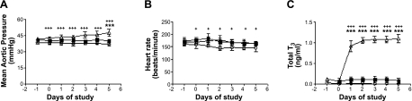
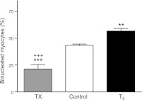
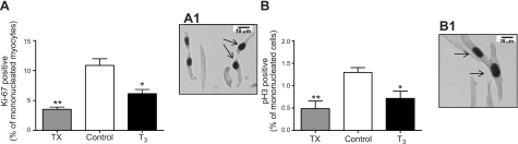
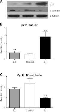
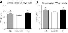
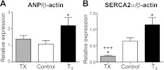
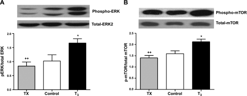
Tri-iodo-l-thyronine (T(3)) suppresses the proliferation of near-term serum-stimulated fetal ovine cardiomyocytes in vitro. Thus, we hypothesized that T(3) is a major stimulant of cardiomyocyte maturation in vivo. We studied 3 groups of sheep fetuses on gestational days 125-130 (term ∼145 d): a T(3)-infusion group, to mimic fetal term levels (plasma T(3) levels increased from ∼0.1 to ∼1.0 ng/ml; t(1/2)∼24 h); a thyroidectomized group, to produce low thyroid hormone levels; and a vehicle-infusion group, to serve as intact controls. At 130 d of gestation, sections of left ventricular freewall were harvested, and the remaining myocardium was enzymatically dissociated. Proteins involved in cell cycle regulation (p21, cyclin D1), proliferation (ERK), and hypertrophy (mTOR) were measured in left ventricular tissue. Evidence that elevated T(3) augmented the maturation rate of cardiomyocytes included 14% increased width, 31% increase in binucleation, 39% reduction in proliferation, 150% reduction in cyclin D1 protein, and 500% increase in p21 protein. Increased expression of phospho-mTOR, ANP, and SERCA2a also suggests that T(3) promotes maturation and hypertrophy of fetal cardiomyocytes. Thyroidectomized fetuses had reduced cell cycle activity and binucleation. These findings support the hypothesis that T(3) is a prime driver of prenatal cardiomyocyte maturation.


Figures

Figure 1.
Figure 1.
Hemodynamic measurements with changes in thyroid status. A) Mean aortic pressure did not differ between control (solid squares) and TX (open circles), but pressure was significantly increased on d 5 in T3-infused fetuses (open triangles). Mean aortic pressure was depressed in TX compared to T3-infused fetuses throughout the study period. B) Heart rate was decreased significantly in the TX group. C) Total T3 levels were unchanged in control fetuses, while T3 infusion increased concentrations to those found near term. T3 was undetectable in TX fetuses. Data are means ± se; n = 8/group. ***P < 0.001 vs. control; +P < 0.05, +++P < 0.001 vs. TX.
Figure 2.
Figure 2.
Increased circulating T3 promotes terminal differentiation of fetal sheep cardiomyocytes. Elevated T3 concentration increased the portion of LV myocytes that were binucleated. Data are means ± se; n = 8/group. **P < 0.01, ***P < 0.001 vs. control; +++P < 0.001 vs. T3.
Figure 3.
Figure 3.
A) Cell cycle activity, measured by Ki-67, decreased in both T3-infused and TX fetuses. A1) Image at ×400. B) Mitotic activity of myocytes, measured by phospho-histone 3 expression, was decreased in both T3-infused and TX fetuses. B1) Image at ×400. Arrows indicate positively labeled cardiomyocytes. Data are means ± se; n = 8/group. *P < 0.05; **P < 0.01 vs. control.
Figure 4.
Figure 4.
A) Immunoblots of cell cycle proteins in cardiomyocytes from T3-infused and TX fetuses compared to controls. B) Cell cycle suppressant p21 was markedly elevated in T3-infused compared to control and TX fetuses. C) Expression of cell cycle promoter cyclin D1 is diminished in T3-infused compared to control and TX fetuses. Data are means ± se; n = 8/group. **P < 0.05 vs. control; ++P < 0.01 vs. T3.
Figure 5.
Figure 5.
T3 promotes cardiomyocyte hypertrophy. Binucleated cardiomyocyte width was increased in left (A) and right (B) ventricles from fetuses that were either deficient or had elevated circulating T3. Both elevated T3 levels and T3 deprivation led to widening of ventricular binucleated myocytes. Data are means ± se; n = 8/group. *P < 0.05, ***P < 0.001 vs. control; +P < 0.05 vs. T3.
Figure 6.
Figure 6.
T3 promotes molecular markers of cardiomyocyte maturation. A) Expression of ANP was increased in the left ventricles of fetuses receiving exogenous T3, but thyroidectomy did not alter ventricular ANP. B) SERCA2a expression levels were lower in TX fetal hearts and higher in T3-infused hearts compared to controls. Data are means ± se; n = 8/group. *P < 0.05 vs. control; +++P < 0.001 vs. T3.
Figure 7.
Figure 7.
T3 activates ERK and mTOR. Immunoblots showed elevated levels of phosphorylated ERK (A) and mTOR (B) in fetal hearts exposed to elevated levels T3, but phosphoprotein levels were unchanged in hearts from TX fetuses. Data are means ± se; n = 8/group. *P < 0.05 vs. control; ++P < 0.01 vs. T3.

Similar articles

Cited by

References

    1. Brown D. D., Cai L. (2007) Amphibian metamorphosis. Dev. Biol. 306, 20–33 - PMC - PubMed
    1. Breall J. A., Rudolph A. M., Heymann M. A. (1984) Role of thyroid hormone in postnatal circulatory and metabolic adjustments. J. Clin. Invest. 73, 1418–1424 - PMC - PubMed
    1. Fisher D. A. (1997) Fetal thyroid function: diagnosis and management of fetal thyroid disorders. Clin. Obstet. Gynecol. 40, 16–31 - PubMed
    1. Macchia P. E. (2000) Recent advances in understanding the molecular basis of primary congenital hypothyroidism. Mol. Med. Today. 6, 36–42 - PubMed
    1. Bianco A. C., Kim B. W. (2006) Deiodinases: implications of the local control of thyroid hormone action. J. Clin. Invest. 116, 2571–2579 - PMC - PubMed

Publication types

MeSH terms