Review
. 2012 Feb;59(2):284-92.
doi: 10.1053/j.ajkd.2011.07.024. Epub 2011 Oct 5.

New insights into human minimal change disease: lessons from animal models

Affiliations
Review

New insights into human minimal change disease: lessons from animal models

Sumant S Chugh et al. Am J Kidney Dis. 2012 Feb.

Abstract

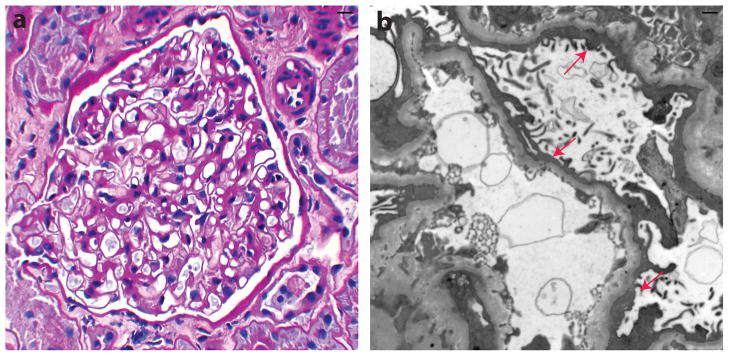
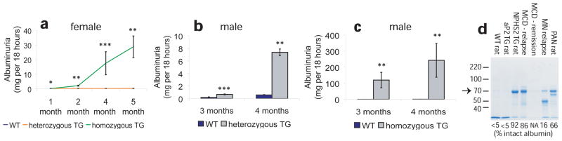
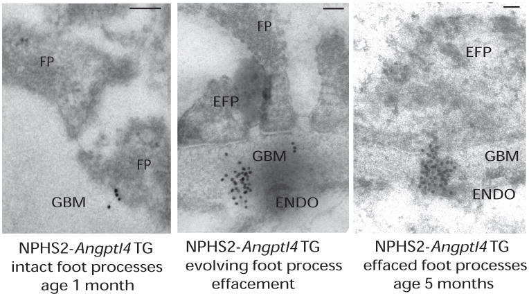
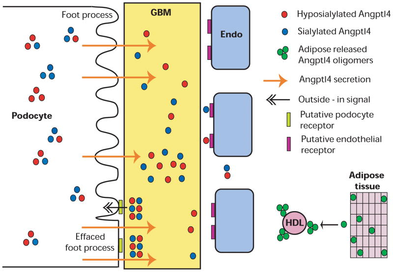
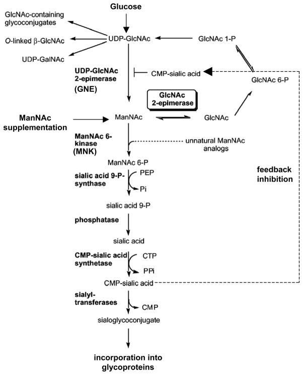
The pathogenesis of minimal change disease (MCD), considered to be the simplest form of nephrotic syndrome, has been one of the major unsolved mysteries in kidney disease. In this review, recent landmark studies that have led to the unraveling of MCD are discussed. A recent study now explains the molecular basis of major clinical and morphologic changes in MCD. Overproduction of angiopoietin-like 4 (ANGPTL4) in podocytes in MCD causes binding of ANGPTL4 to the glomerular basement membrane, development of nephrotic-range selective proteinuria, diffuse effacement of foot processes, and loss of glomerular basement membrane charge, but is not associated with changes shown by light microscopy in the glomerular and tubulointerstitial compartments. At least some of this ability of ANGPTL4 to induce proteinuria is linked to a deficiency of sialic acid residues because oral supplementation with sialic acid precursor N-acetyl-d-mannosamine improves sialylation of podocyte-secreted ANGPTL4 and significantly decreases proteinuria. Animal models of MCD, recent advances in potential biomarkers, and studies of upstream factors that may initiate glomerular changes also are discussed. In summary, recent progress in understanding MCD is likely to influence the diagnosis and treatment of MCD in the near future.


Figures

Figure 1
Figure 1
Histological characteristics of human minimal change disease. (a) Periodic acid–Schiff stain shows normal appearance of glomeruli by light microscopy. (b) Electron microscopy reveals diffuse effacement of podocyte foot processes (arrows). Courtesy of Dr William Cook.
Figure 2
Figure 2
Relationship of ANGPTL4 overexpression with proteinuria. (a) Albuminuria in female NPHS2-ANGPTL4 transgenic rats. (b) Albuminuria in male heterozygous NPHS2-ANGPTL4 transgenic rats. (c) Albuminuria in male homozygous NPHS2-ANGPTL4 transgenic rats. (d) Gel electrophoresis of urinary protein from wild-type (WT) rats, transgenic (TG) rats, PAN (puromycin aminonucleoside nephrosis) rats, and individuals with minimal change disease (MCD) and membranous nephropathy (MN). Arrow points towards prominent 70 kDa intact albumin band. Mean percentage densitometry of intact albumin is shown for each lane. * P<0.05, ** P<0.01, *** P<0.001. Abbreviations: NPHS2, podocin; ANGPTL4, angiopoietin-like 4. Reproduced from Clement et al.
Figure 3
Figure 3
Relationship of clustering of ANGPTL4 with effacement of podocyte foot processes. Immunogold electron microscopy of NPHS2-ANGPTL4 transgenic rats revealed a progression from (left) intact foot processes (FP) with gold particles just entering the glomerular basement membrane (GBM) in young rats, (middle) to partial effacement with GBM gold particle clusters opposite to effaced foot processes (EFP) reaching up to the endothelial (ENDO) surface (usually noted starting age 3 months), and finally (right) diffuse effacement with dense gold particle clusters in the GBM usually noted by age 5 months. Scale bars denote 0.2 μm. Abbreviation: TG, transgenic. Reproduced from Clement et al.
Figure 4
Figure 4
Schematic representation of the role of podocyte secreted Angtpl4 in the pathogenesis of minimal change disease. Sequence of events arranged from top to bottom. Podocytes secrete neutral and high pI ANGPTL4 that binds to the glomerular basement membrane (GBM) to alter protein-protein interactions, resulting in proteinuria. Over time, ANGPTL4 reaches up to the endothelial (Endo) surface. Progressive accumulation and clustering of ANGPTL4 in the GBM likely activates signals at the podocyte-GBM interface and induces foot process effacement. Circulating ANGPTL4 secreted from other organs in disease states, e. g. adipose tissue, forms medium and high order oligomers that are bound to high-density lipoprotein (HDL) particles, migrate at neutral or low-neutral pI, and do not enter the GBM or cause proteinuria. Reproduced from Clement et al.
Figure 5
Figure 5
Brief schematic representation of the sialic acid biosynthesis pathway. Glucose is converted into uridine diphosphate–N-acetylglucosamine (UDP-GlcNAc) through a number of enzymatic steps (not shown), which is converted to N-acetyl-D-mannosamine (ManNAc) via a rate-limiting reaction calalyzed by GNE. ManNAc undergoes conversion to cytidine monophosphate–sialic acid (CMP-sialic acid), which provides feedback inhibition to the rate-limiting GNE-calalyzed reaction. Exogenous ManNAc supplements enters the pathway after this rate limiting step. A substantial part of the sialic acid is incorporated into N- and O-linked glycan residues in structural and secreted glycoproteins. Modified from Luchansky et al with permission of The American Society for Biochemistry and Molecular Biology.

Similar articles

Cited by

References

    1. Nachman PH, Jennette JC, Falk R. Primary glomerular disease. In: Brenner BM, editor. The Kidney. 8. Philadelphia, PA: Elsevier; 2008. pp. 987–1066.
    1. Miner JH. Glomerular filtration: the charge debate charges ahead. Kidney Int. 2008;74(3):259–261. - PubMed
    1. Pippin JW, Brinkkoetter PT, Cormack-Aboud FC, et al. Inducible rodent models of acquired podocyte diseases. Am J Physiol Renal Physiol. 2009;296(2):F213–F229. - PubMed
    1. Seiler MW, Venkatachalam MA, Cotran RS. Glomerular epithelium: structural alterations induced by polycations. Science. 1975;189(4200):390–393. - PubMed
    1. Seiler MW, Rennke HG, Venkatachalam MA, Cotran RS. Pathogenesis of polycation-induced alterations (“fusion”) of glomerular epithelium. Lab Invest. 1977;36(1):48–61. - PubMed