. 2011 Oct 5:11:90.
doi: 10.1186/1472-6750-11-90.

Design and development of a peptide-based adiponectin receptor agonist for cancer treatment

Affiliations

Design and development of a peptide-based adiponectin receptor agonist for cancer treatment

Laszlo Otvos Jr et al. BMC Biotechnol. .

Abstract

Background: Adiponectin, a fat tissue-derived adipokine, exhibits beneficial effects against insulin resistance, cardiovascular disease, inflammatory conditions, and cancer. Circulating adiponectin levels are decreased in obese individuals, and this feature correlates with increased risk of developing several metabolic, immunological and neoplastic diseases. Thus, pharmacological replacement of adiponectin might prove clinically beneficial, especially for the obese patient population. At present, adiponectin-based therapeutics are not available, partly due to yet unclear structure/function relationships of the cytokine and difficulties in converting the full size adiponectin protein into a viable drug.

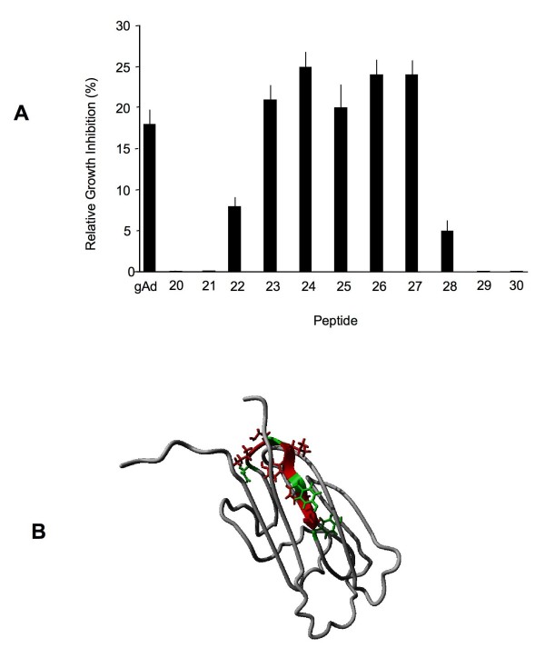
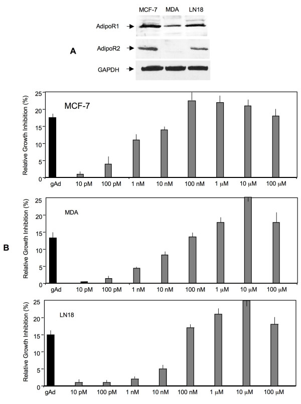
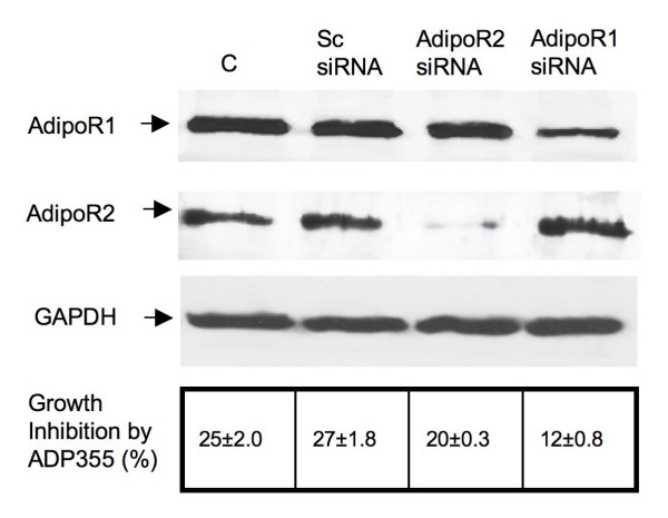
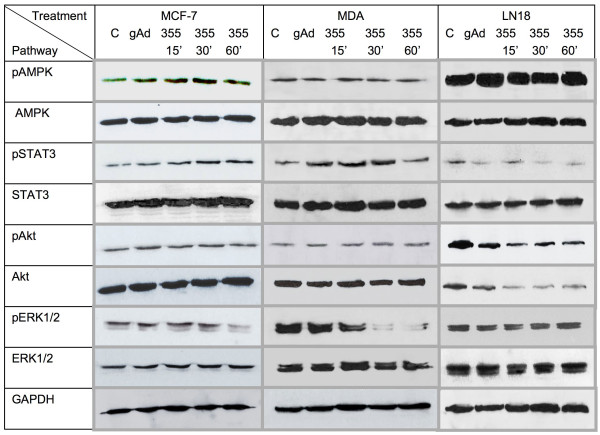
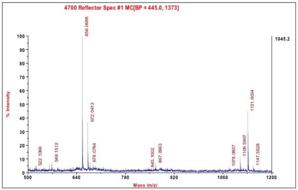
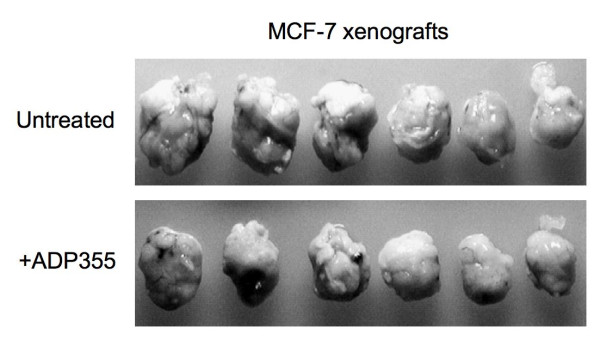
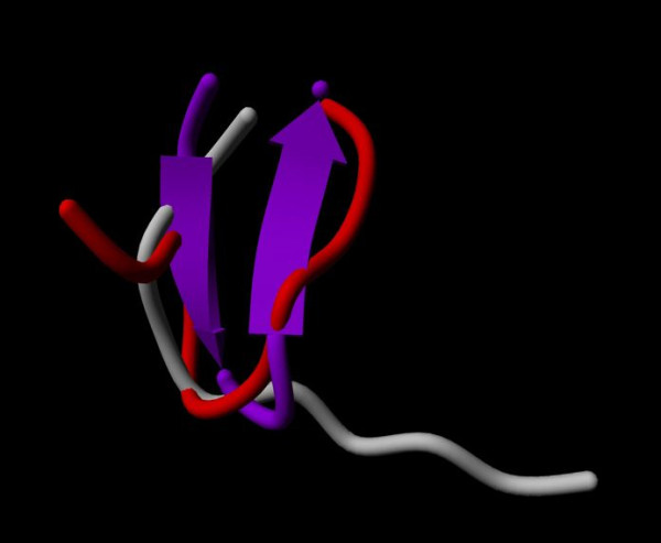
Results: We aimed to generate adiponectin-based short peptide that can mimic adiponectin action and be suitable for preclinical and clinical development as a cancer therapeutic. Using a panel of 66 overlapping 10 amino acid-long peptides covering the entire adiponectin globular domain (residues 105-254), we identified the 149-166 region as the adiponectin active site. Three-dimensional modeling of the active site and functional screening of additional 330 peptide analogs covering this region resulted in the development of a lead peptidomimetic, ADP 355 (H-DAsn-Ile-Pro-Nva-Leu-Tyr-DSer-Phe-Ala-DSer-NH2). In several adiponectin receptor-positive cancer cell lines, ADP 355 restricted proliferation in a dose-dependent manner at 100 nM-10 μM concentrations (exceeding the effects of 50 ng/mL globular adiponectin). Furthermore, ADP 355 modulated several key signaling pathways (AMPK, Akt, STAT3, ERK1/2) in an adiponectin-like manner. siRNA knockdown experiments suggested that ADP 355 effects can be transmitted through both adiponectin receptors, with a greater contribution of AdipoR1. In vivo, intraperitoneal administration of 1 mg/kg/day ADP 355 for 28 days suppressed the growth of orthotopic human breast cancer xenografts by ~31%. The peptide displayed excellent stability (at least 30 min) in mouse blood or serum and did not induce gross toxic effects at 5-50 mg/kg bolus doses in normal CBA/J mice.

Conclusions: ADP 355 is a first-in-class adiponectin receptor agonist. Its biological activity, superior stability in biological fluids as well as acceptable toxicity profile indicate that the peptidomimetic represents a true lead compound for pharmaceutical development to replace low adiponectin levels in cancer and other malignancies.


Figures

Figure 1
Figure 1
Identification of the active site of adiponectin. A) Effects of adiponectin fragments encompassing the active site on the growth of MCF7 cells. The activity of the entire globular domain of adiponectin (gAd) is included for comparison. The data are averages from 3 different assays and represent average results +/- SE and were analyzed by Student t-test, p < 0.05. The sequences of all tested peptides are listed in Additional file 1. B) High-resolution structure of the adiponectin monomer with the peptide 25 and active site amino acid side-chains colored. Conservative substitutions of residues marked in green could be made without loss of biological activity; residues marked with red could not be substituted.
Figure 2
Figure 2
Effects of ADP 355 on the growth of cancer cells in vitro. A) Expression of AdipoR1 (49 kDa) and AdipoR2 (44 kDa) in MCF-7, MDA-MB-231 and LN18 cells was examined by WB, as described in Methods. B) Cytostatic activity of ADP 355 at 10-100 μM was assessed in MCF-7, MDA-MB-231, and LN18 cancer cell lines, as described in Methods. Bars represent % growth inhibition relative to untreated cells +/- SE.
Figure 3
Figure 3
Effects of siRNA-mediated downregulation of AdipoR1 or AdipoR2 on ADP 355 activity. The expression of AdipoR1, AdipoR2, and control protein GAPDH were assessed by WB in control (C) cells (transfection medium only), cells treated with scrambled siRNA (Sc siRNA), siRNA targeting AdipoR1, or siRNA targeting AdipoR2, as described in Methods. The relative levels of AdipoR1 and AdipoR2 proteins were calculated by densitometry scanning, as described in Methods, and are provided in Additional file 2. The relative % of growth inhibition upon ADP 355 treatment in cells with different levels of AdipoR1 and AdipoR2 vs. untreated cells is shown in the lower panel table.
Figure 4
Figure 4
Effects of ADP 355 on intracellular cell signaling in cancer cells. The effects of ADP 355 on signaling pathways in MCF-7, MDA-MB-231, and LN18 cells at 0-60 min of treatment were studied by WB, as described in Methods. The expression of GAPDH was used as determination of protein loading. The relative levels of phosphorylated/total proteins were calculated by densitometry scanning, as described in Methods, and are provided in Additional file 3.
Figure 5
Figure 5
Stability of ADP 355 in whole mouse blood. The peptide stability was assessed in whole mouse blood after 30 min of incubation by mass spectroscopy as described in Methods. The only peptide-originated peaks are at 1109 and 1131 M/z, representing the unmodified peptide and its sodium adduct.
Figure 6
Figure 6
Anti-tumor ADP 355 activity in vivo. Orthotopic MCF-7 xenografts were established as described in Methods. After 34 days, 5 mice were treated with ADP 355 at 1 mg/kg/day dose, and 5 mice remained untreated. After 28 days, the mice were sacrificed and the lesions removed. Due to variability in tumor sizes, the largest and smallest lesions from each group were excluded from the evaluation. The excised, middle-sized lesions, from 3 treated and 3 untreated mice are shown.
Figure 7
Figure 7
ADP 355 energy analysis. Representative energy minimized structures of peptide 25 (red) and ADP 355 (purple) overlaid to the conformation of the 153-162 sequence found in adiponectin protein (grey).

Similar articles

Cited by

References

    1. Grossmann ME, Nkhata KJ, Mizuno NK, Ray A, Cleary MP. Effects of adiponectin on breast cancer cell growth and signaling. Br J Cancer. 2008;98(2):370–379. doi: 10.1038/sj.bjc.6604166. - DOI - PMC - PubMed
    1. Ryan AS, Berman DM, Nicklas BJ, Sinha M, Gingerich RL, Meneilly GS, Egan JM, Elahi D. Plasma adiponectin and leptin levels, body composition, and glucose utilization in adult women with wide ranges of age and obesity. Diabetes Care. 2003;26(8):2383–2388. doi: 10.2337/diacare.26.8.2383. - DOI - PubMed
    1. Chiarugi P, Fiaschi T. Adiponectin in health and diseases: from metabolic syndrome to tissue regeneration. Expert Opin Ther Targets. 2010;14(2):193–206. doi: 10.1517/14728220903530712. - DOI - PubMed
    1. Chen X, Wang Y. Adiponectin and breast cancer. Med Oncol. 2010. - PubMed
    1. Shibata R, Ouchi N, Murohara T. Adiponectin and cardiovascular disease. Circ J. 2009;73(4):608–614. doi: 10.1253/circj.CJ-09-0057. - DOI - PubMed

Publication types

MeSH terms