. 2012 Sep;72(9):1181-95.
doi: 10.1002/dneu.20984. Epub 2012 Jun 21.

Hindered submicron mobility and long-term storage of presynaptic dense-core granules revealed by single-particle tracking

Affiliations

Hindered submicron mobility and long-term storage of presynaptic dense-core granules revealed by single-particle tracking

B A Scalettar et al. Dev Neurobiol. 2012 Sep.

Abstract

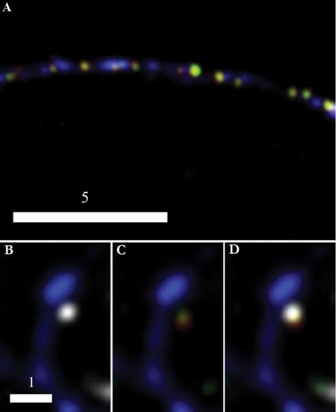
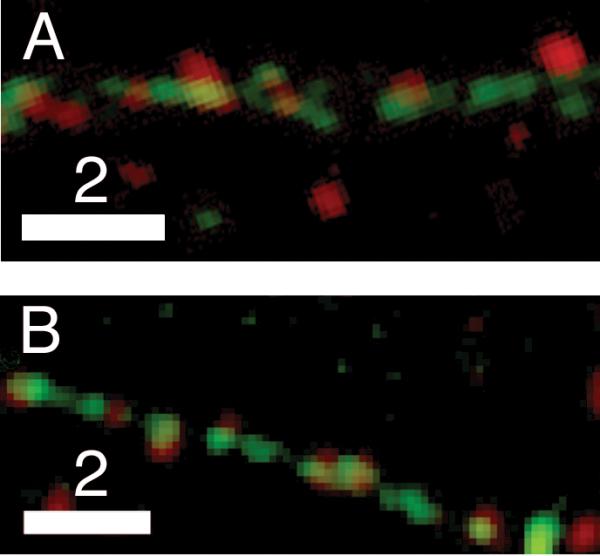
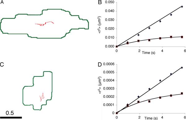
Dense-core granules (DCGs) are organelles found in neuroendocrine cells and neurons that house, transport, and release a number of important peptides and proteins. In neurons, DCG cargo can include the secreted neuromodulatory proteins tissue plasminogen activator (tPA) and/or brain-derived neurotrophic factor (BDNF), which play a key role in modulating synaptic efficacy in the hippocampus. This function has spurred interest in DCGs that localize to synaptic contacts between hippocampal neurons, and several studies recently have established that DCGs localize to, and undergo regulated exocytosis from, postsynaptic sites. To complement this work, we have studied presynaptically localized DCGs in hippocampal neurons, which are much more poorly understood than their postsynaptic analogs. Moreover, to enhance relevance, we visualized DCGs via fluorescence labeling of exogenous and endogenous tPA and BDNF. Using single-particle tracking, we determined trajectories of more than 150 presynaptically localized DCGs. These trajectories reveal that mobility of DCGs in presynaptic boutons is highly hindered and that storage is long-lived. We also computed mean-squared displacement curves, which can be used to elucidate mechanisms of transport. Over shorter time windows, most curves are linear, demonstrating that DCG transport in boutons is driven predominantly by diffusion. The remaining curves plateau with time, consistent with motion constrained by a submicron-sized corral. These results have relevance to recent models of presynaptic organization and to recent hypotheses about DCG cargo function. The results also provide estimates for transit times to the presynaptic plasma membrane that are consistent with measured times for onset of neurotrophin release from synaptically localized DCGs.


Figures

Fig. 1
Fig. 1
DCGs containing tPA or BDNF localize to presynaptic sites. Representative deblurred three-color fluorescence image (A) of dendritic processes (green), SV clusters (blue) present in axons of their synaptic partners, and tPA-containing DCGs (red). The boxed region in (A) is enlarged in (B), and several tPA-containing DCGs that colocalize with presynaptic sites are identified with arrowheads. Analogous images (C-F) from hippocampal neurons expressing BDNF-mCherry. BDNF-containing DCGs that colocalize with presynaptic sites are identified with arrowheads. The lowest arrowhead in (C) points to a DCG localized to a shaft synapse. Scale Bar = 10 μm for A. Scale Bar = 2 μm for B-F.
Fig. 2
Fig. 2
BDNF-mCherry and tPA-EYFP are copackaged in presynaptic DCGs. Deblurred fluorescence image (A) of an axon from a 14-DIV hippocampal neuron showing colocalization of tPA-EYFP (green) and BDNF-mCherry (red) within DCGs that, in turn, colocalize with SV clusters (blue) immunostained against synapsin-1 using a Cascade Blue secondary. DCGs containing tPA and BDNF appear yellow. Images (B-D) of a synapse between fixed 21-DIV hippocampal neurons. DCG proteins are tagged as in (A) and the SV cluster (white) again is immunostained against synapsin-1, but with a secondary, Alexa Fluor 647, that emits in the far red. The spine (blue) is labeled using an EBFP2 fill. For clarity, the spine is shown first with the SV cluster (B) and then with the two overlapping DCG proteins (C). All stained components are shown together in (D). Scale Bar = 5 μm for A. Scale Bar = 1 μm for B-D.
Fig. 3
Fig. 3
Endogenous neuromodulators localize to presynaptic sites in hippocampal neurons. Dual-color image showing that puncta stained by an antibody against endogenous BDNF (red) (A) are present in presynaptic clusters (green) along axons of fixed 7-DIV hippocampal neurons expressing synaptophysin-EGFP. Analogous image of endogenous tPA (red) (B) in fixed 10-DIV hippocampal neurons expressing synaptophysin-EGFP (green). Quantification of data obtained from these types of images revealed that the probability that a bouton houses a DCG containing endogenous BDNF is 0.41 ± 0.04 (n = 171 SV clusters, 65 DCGs). The analogous result for endogenous tPA is 0.39 ± 0.07 (n = 382 SV clusters, 171 DCGs). Each of these endogenous localization probabilities is statistically indistinguishable from its analogous exogenous localization probability. Specifically, results from the Student's t-test yielded large p values for both BDNF (p = 0.40 > 0.05) and tPA (p = 0.49 > 0.05). Scale Bar = 2 μm for A and B. In past work, several groups (including ours) also have used immunostaining to compare levels of expression of endogenous and exogenous neuromodulators. These analyses reveal that, on average, exogenous neuromodulators are expressed at levels that are ~2–5× endogenous levels (Kolarow et al., 2007, Lochner et al., 2008, Matsuda et al., 2009) and that the density of DCGs containing exogenous neuromodulators is less than twice that of DCGs containing endogenous neuromodulators. We tend to find exogenous protein chimeras expressed at the lower end of this range, perhaps because we image neurons quickly after viral delivery of DNAs encoding the chimeras. The important conclusion from these studies is that overexpression of chimeras is modest and thus is unlikely to introduce major artifacts into our results, such as extensive labeling of DCGs that do not contain endogenous neuromodulators.
Fig. 4
Fig. 4
DCGs at presynaptic sites undergo very slow Brownian diffusion and corralled motion. Trajectory (A) of a relatively mobile presynaptic DCG and associated plots (B) of <r2> versus t deduced from two relatively mobile presynaptic DCGs. One plot (◆) is linear, consistent with Brownian diffusion, and one (■) is nonlinear, consistent with corralled motion. Trajectory (C) and associated plots (D) of <r2> versus t deduced from nearly immobile presynaptic DCGs. In panels B and D, the Brownian data were divided by 10 to facilitate displaying two data sets on the same graph. In panels A and C, DCGs are represented by points localized at their center of mass and thus do not extend over a representative fraction of the cluster. Scale bar = 0.5 μm.
Fig. 5
Fig. 5
DCGs remain localized at presynaptic sites for a few hours. Two-color image (A) of 17-DIV hippocampal neurons expressing BDNF-mCherry (red) and synaptophysin-EGFP (green). A DCG that colocalized with an SV cluster for at least 90 minutes is identified with an arrowhead, and the trajectory of the DCG for a 15-minute time window is shown in blue, slightly offset from the SV cluster. Plot of <r2> versus t (B) obtained from tracking the DCG in panel A for 15 minutes. The line in panel B is a fit to the corralled diffusion model. Scale bar = 2.0 μm. Supplemental Movie 1. Visualization of DCG transport in the soma, along processes, and in boutons of mature neurons. Deblurred time-lapse movie showing transport of DCGs in a 17-DIV hippocampal neuron expressing synaptophysin-EGFP (green) and BDNF-mCherry (red).

References

    1. Abney JR, Meliza CD, Cutler B, Kingma M, Lochner JE, Scalettar BA. Real-time imaging of the dynamics of secretory granules in growth cones. Biophys J. 1999;77:2887–2895. - PMC - PubMed
    1. Barg S, Olofsson CS, Schriever-Abeln J, Wendt A, Gebre-Medhin S, Renstrom E, Rorsman P. Delay between fusion pore opening and peptide release from large dense-core vesicles in neuroendocrine cells. Neuron. 2002;33:287–299. - PubMed
    1. Benchenane K, Lopez-Atalaya JP, Fernandez-Monreal M, Touzani O, Vivien D. Equivocal roles of tissue-type plasminogen activator in stroke-induced injury. Trends Neurosci. 2004;27:155–160. - PubMed
    1. Benson DL, Salton SR. Expression and polarization of VGF in developing hippocampal neurons. Brain Res Dev Brain Res. 1996;96:219–228. - PubMed
    1. Berezhkovskii AM, Grigoriev IV, Makhnovskii YA, Zitserman VY. Kinetics of escape through a small hole. Journal of Chemical Physics. 2002;116:9574–9577.

Publication types

Substances