Comparative Study
. 2011 Oct 5;31(40):14264-71.
doi: 10.1523/JNEUROSCI.2310-11.2011.

NBCe1 mediates the acute stimulation of astrocytic glycolysis by extracellular K+

Affiliations
Comparative Study

NBCe1 mediates the acute stimulation of astrocytic glycolysis by extracellular K+

Iván Ruminot et al. J Neurosci. .

Abstract

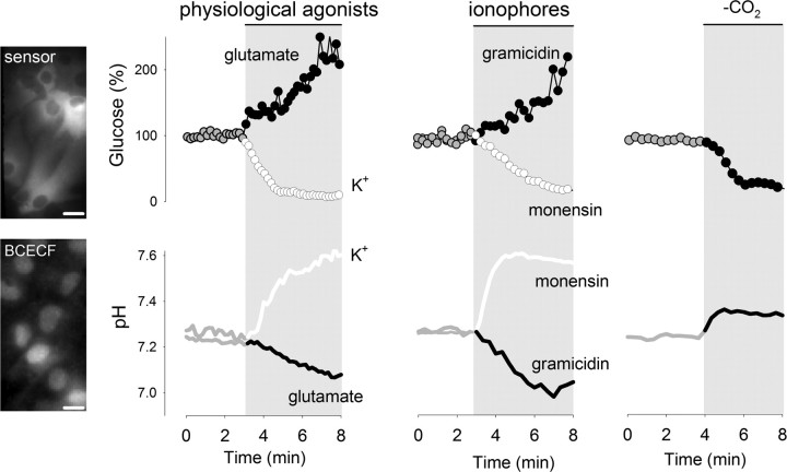
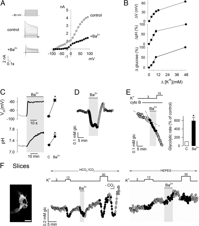
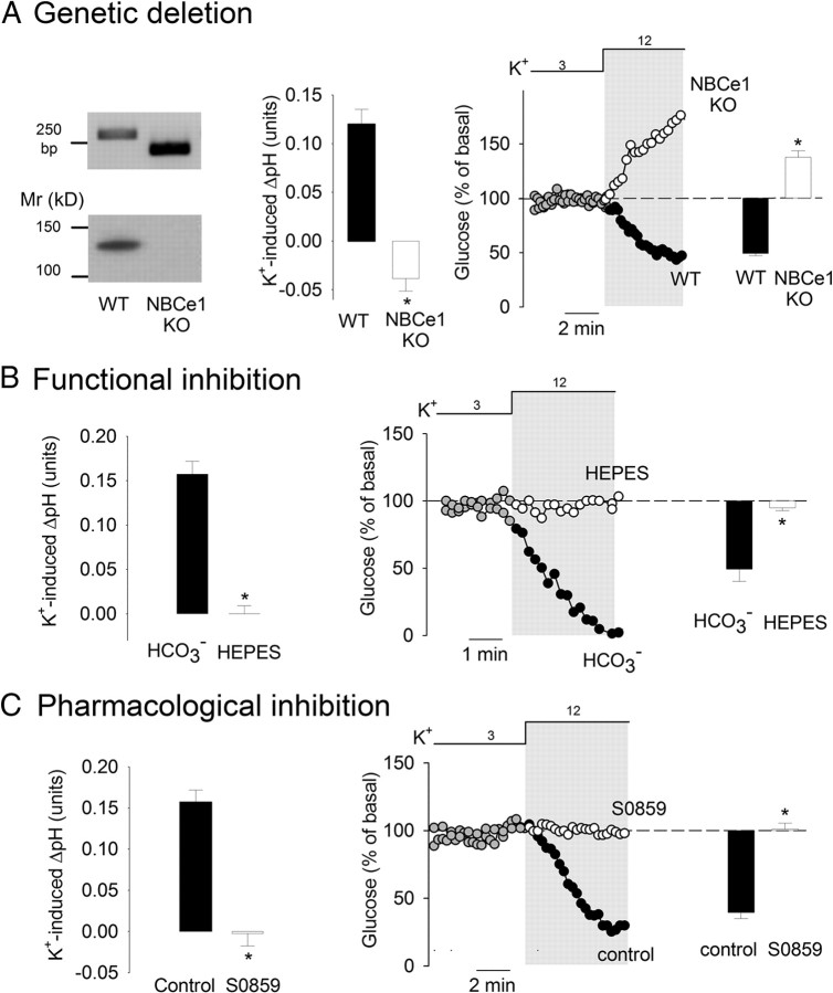
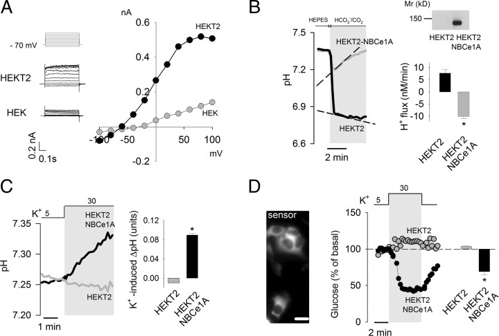
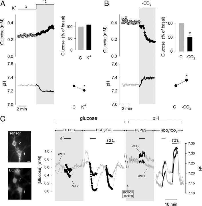
Excitatory synaptic transmission stimulates brain tissue glycolysis. This phenomenon is the signal detected in FDG-PET imaging and, through enhanced lactate production, is also thought to contribute to the fMRI signal. Using a method based on Förster resonance energy transfer in mouse astrocytes, we have recently observed that a small rise in extracellular K(+) can stimulate glycolysis by >300% within seconds. The K(+) response was blocked by ouabain, but intracellular engagement of the Na(+)/K(+) ATPase pump with Na(+) was ineffective, suggesting that the canonical feedback regulatory pathway involving the Na(+) pump and ATP depletion is only permissive and that a second mechanism is involved. Because of their predominant K(+) permeability and high expression of the electrogenic Na(+)/HCO(3)(-) cotransporter NBCe1, astrocytes respond to a rise in extracellular K(+) with plasma membrane depolarization and intracellular alkalinization. In the present article, we show that a fast glycolytic response can be elicited independently of K(+) by plasma membrane depolarization or by intracellular alkalinization. The glycolytic response to K(+) was absent in astrocytes from NBCe1 null mice (Slc4a4) and was blocked by functional or pharmacological inhibition of the NBCe1. Hippocampal neurons acquired K(+)-sensitive glycolysis upon heterologous NBCe1 expression. The phenomenon could also be reconstituted in HEK293 cells by coexpression of the NBCe1 and a constitutively open K(+) channel. We conclude that the NBCe1 is a key element in a feedforward mechanism linking excitatory synaptic transmission to fast modulation of glycolysis in astrocytes.


Figures

Figure 1.
Figure 1.
Acute modulation of astrocytic glycolysis by intracellular pH. Images on the left show astrocytes expressing the glucose sensor FLII12Pglu600μΔ6 or loaded with BCECF, bars represent 10 μm. The effects of K+ (12 mm), glutamate (50 μm), monensin (100 μm), and gramicidin (20 μg/ml) on intracellular glucose concentration and pH were measured as described in Materials and Methods. Intracellular glucose and pH were measured during transition between HCO3 buffers of equal pH, with and without 5% CO2. All traces correspond to representative plots of at least three independent experiments.
Figure 2.
Figure 2.
Acute modulation of astrocytic glycolysis by membrane depolarization. A, The current/voltage relationship in cortical astrocytes was registered by patch clamping as described in Materials and Methods. Currents were recorded from a holding potential of −80 mV at variable 500 ms test pulses from −100 to +100 mV, in the absence and presence of 3 mm Ba2+. B, Effect of increasing K+ concentrations on membrane potential (n = 3 cells), pH (n = 3), and glucose concentration (n = 9 cells). Traces are representative of three independent experiments. Initial rates of pH increase and glucose decrease were estimated over 3 min. C, Membrane potential and intracellular pH was measured before and after exposure to 3 mm Ba2+. Bars summarize data from three experiments. D, Effect of 3 mm Ba2+ on intracellular glucose in a single astrocyte, representative of three similar experiments. E, Sequential effects of 3 mm Ba2+ and 15 mm K+ on the glycolytic rate of a single astrocyte. Bars summarize data from three experiments. F, Image on the left shows a 3D confocal reconstruction of two astrocytes expressing the glucose sensor FLII12Pglu600μΔ6 in an organotypical hippocampal slice. Bar represents 10 μm. Graphs illustrate experiments in slices testing the effects of 12 mm K+ and 3 mm Ba2+ on intracellular glucose in the presence and absence (HEPES) of HCO3. Data are representative of three separate experiments.
Figure 3.
Figure 3.
NBCe1 is necessary for K+-stimulated glycolysis in astrocytes. A, Genetic deletion of NBCe1. The left panel illustrates the results of PCR genotyping of tail biopsies and Western blotting analysis of cultured astrocytes from wild-type (WT) and NBCe1 knock-out mice (KO). Bars in the middle represent the changes in intracellular pH elicited by a 3 min exposure to 12 mm K+ in WT (n = 3) and NBCe1 KO astrocytes (n = 7). The impact of a 3 min exposure to 12 mm K+ on glucose concentration in WT and NBCe1 KO astrocytes is illustrated by the time course and bar graph on the right (n = 3–4). B, Bicarbonate omission. Bars on the left illustrate pH changes elicited by a 3 min exposure to 12 mm K+ in the presence and absence (HEPES) of bicarbonate (n = 4). Middle, The effect of 12 mm K+ on glucose concentration was measured over time in the presence and absence (HEPES) of bicarbonate. Bars on the right show the change in intracellular glucose after 3 min (n = 3). C, S0859. Bars on the left illustrate pH changes elicited by a 3 min exposure to 12 mm K+ before and after a 4 min preincubation with 30 μm S0859 (n = 3). Middle, The effect of 12 mm K+ on glucose concentration was measured before and after a 4 min preincubation with 30 μm S0859 (n = 3). Bars on the right show the change in intracellular glucose after 3 min (n = 3).
Figure 4.
Figure 4.
Assembly of K+-modulated glycolysis in HEK cells. A, The steady-state current/voltage relationship in HEK and HEKT2 cells was measured by patch clamp as described in Materials and Methods. The left panels show the actual currents recorded from a holding potential of −80 mV at different 500 ms test pulses from −100 to +100 mV. B, Protein expression in HEK and HEKT2 cells overexpressing NBCe1A was measured by Western blotting as described in Materials and Methods. NBCe1 activity was assessed by estimating the rate of acid extrusion in response to an acid load induced by switching from HEPES to HCO3/CO2 in the presence of the NHE inhibitor amiloride (1 mm). The bar graph summarizes data from four experiments. C, The effect of K+ rise (from 5 to 30 mm) on intracellular pH was measured in HEKT2 and HEKT2-NBCe1A cells. The bar graph shows the change in pH after 3 min (n = 3). D, HEKT2 cells were cotransfected with cDNAs coding for the FRET glucose sensor and NBCe1A, bar represents 10 μm. The effect of K+ rise (from 5 to 30 mm) on intracellular glucose was measured in HEKT2 and HEKT2-NBCe1A cells. Bars show the change in glucose concentration after 3 min (n = 3).
Figure 5.
Figure 5.
Lack of K+-stimulated glycolysis in hippocampal neurons is explained by absence of NBCe1. A, Time course of the effects of 12 mm K+ on neuronal glucose (top) and pH (bottom). Graphs on the right summarize the changes after 3 min of exposure (n = 3 or 4). B, Time course of the effect of CO2 removal on neuronal glucose (top) and pH (bottom). Changes after 3 min are summarized on the right (n = 3). C, Neurons were cotransfected with cDNAs coding for the FRET glucose sensor and NBCe1A, bars represent 10 μm. After monitoring glucose concentration, cells were AM loaded with BCECF, allowing for a series of pH measurements. The graph presents the effect of 12 mm K+ on glucose and pH in the presence and absence of HCO3/CO2 in two neurons of different morphology. The acute alkalinization observed in response to high K+ demonstrates functional NBCe in these two neurons. Similar results were obtained in six independent experiments.

References

    1. Attwell D, Laughlin SB. An energy budget for signaling in the grey matter of the brain. J Cereb Blood Flow Metab. 2001;21:1133–1145. - PubMed
    1. Azarias G, Perreten H, Lengacher S, Poburko D, Demaurex N, Magistretti PJ, Chatton JY. Glutamate transport decreases mitochondrial pH and modulates oxidative metabolism in astrocytes. J Neurosci. 2011;31:3550–3559. - PMC - PubMed
    1. Barros LF, Deitmer JW. Glucose and lactate supply to the synapse. Brain Res Rev. 2010;63:149–159. - PubMed
    1. Bittner CX, Loaiza A, Ruminot I, Larenas V, Sotelo-Hitschfeld T, Gutiérrez R, Córdova A, Valdebenito R, Frommer WB, Barros LF. High resolution measurement of the glycolytic rate. Front Neuroenergetics. 2010;2:26. - PMC - PubMed
    1. Bittner CX, Valdebenito R, Ruminot I, Loaiza A, Larenas V, Sotelo-Hitschfeld T, Moldenhauer H, San Martín A, Gutiérrez R, Zambrano M, Barros LF. Fast and reversible stimulation of astrocytic glycolysis by K+ and a delayed and persistent effect of glutamate. J Neurosci. 2011;31:4709–4713. - PMC - PubMed

Publication types