Comparative Study
. 2011 Oct 5;31(40):14291-5.
doi: 10.1523/JNEUROSCI.1148-11.2011.

Rapid loss of dendritic HCN channel expression in hippocampal pyramidal neurons following status epilepticus

Affiliations
Comparative Study

Rapid loss of dendritic HCN channel expression in hippocampal pyramidal neurons following status epilepticus

Sangwook Jung et al. J Neurosci. .

Abstract

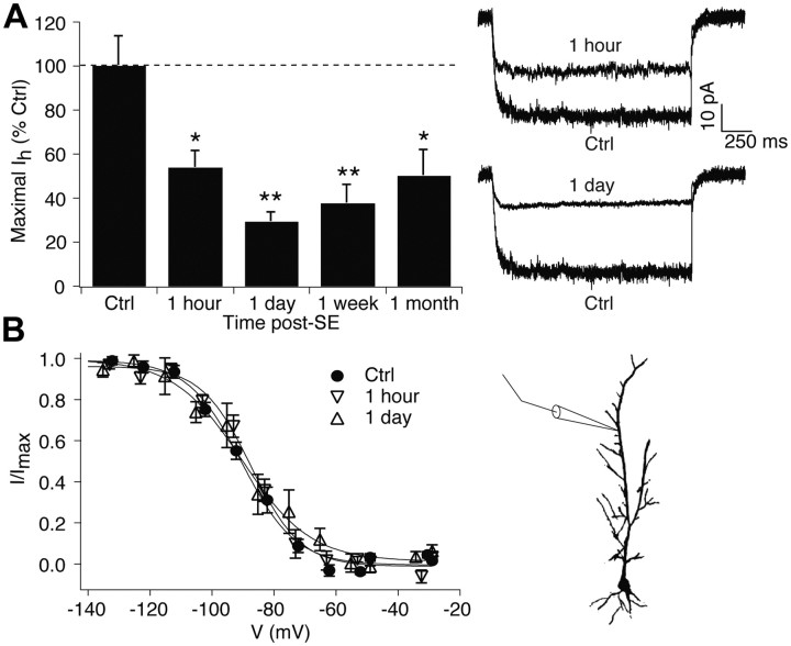
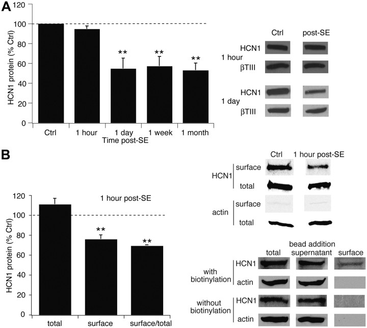
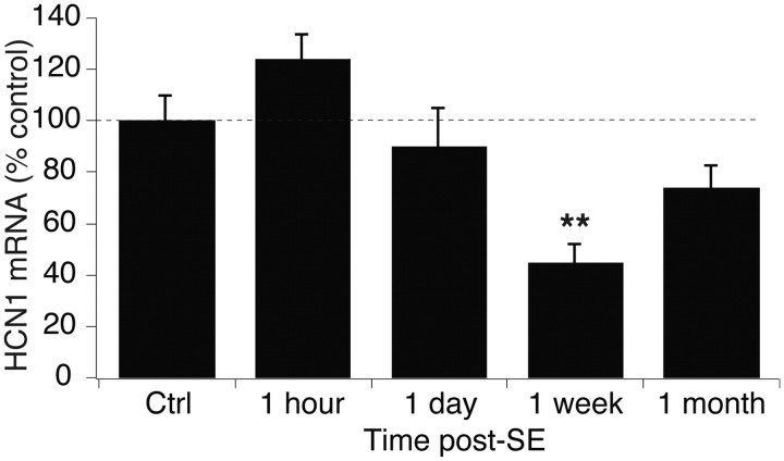
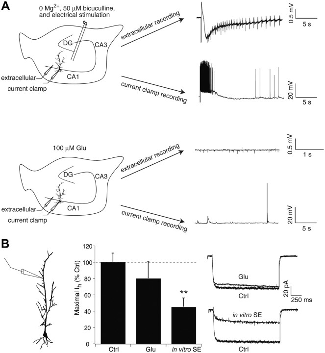
Epilepsy is associated with loss of expression and function of hyperpolarization-activated, cyclic nucleotide-gated (HCN) ion channels. Previously, we showed that loss of HCN channel-mediated current (I(h)) occurred in the dendrites of CA1 hippocampal pyramidal neurons after pilocarpine-induced status epilepticus (SE), accompanied by loss of HCN1 channel protein expression. However, the precise onset and mechanistic basis of HCN1 channel loss post-SE was unclear, particularly whether it preceded the onset of spontaneous recurrent seizures and could contribute to epileptogenesis or development of the epileptic state. Here, we found that loss of I(h) and HCN1 channel expression began within an hour after SE and involved sequential processes of dendritic HCN1 channel internalization, delayed loss of protein expression, and later downregulation of mRNA expression. We also found that an in vitro SE model reproduced the rapid loss of dendritic I(h), demonstrating that this phenomenon was not specific to in vivo SE. Together, these results show that HCN1 channelopathy begins rapidly and persists after SE, involves both transcriptional and nontranscriptional mechanisms, and may be an early contributor to epileptogenesis.


Figures

Figure 1.
Figure 1.
Dendritic Ih loss begins within 1 h after SE and persists for at least 1 month. A, Ih amplitudes at maximal activation obtained from 1 h and 1 d post-SE animals were significantly reduced compared to control. Representative current traces are shown at voltage commands of ∼ −150 mV. Summary data at 1 week are from Jung et al. (2007), and data at 1 month are from Jung et al. (2010). B, Voltage-dependent activation of Ih at 1 h and 1 d post-SE was similar to control. Diagram shows approximate location of dendritic recordings. Ctrl, Control.
Figure 2.
Figure 2.
HCN1 channel protein expression falls after a delay following SE, while surface expression declines immediately at 1 h post-SE. A, HCN1 channel protein expression at 1 h post-SE was unchanged, while at 1 d post-SE was decreased and remained reduced for at least 1 month. Representative blots are shown in each condition, along with blots of β-tubulin III as a marker of neuronal protein content. Summary data at 1 week and 1 month are from Jung et al., (2007). B, Expression of surface HCN1 channel protein was decreased at 1 h post-SE compared to control (Ctrl). Control and post-SE samples were processed in the same gel to enable accurate comparison; total and surface samples were processed in separate gels and thus cannot be directly compared. Lack of actin staining in the surface fraction confirmed the absence of cytoplasmic proteins. Bottom panels from a single representative gel show that no surface protein was recovered with avidin-complexed beads when biotinylation was omitted.
Figure 3.
Figure 3.
HCN1 mRNA levels are reduced within 1 week post-SE. HCN1 mRNA levels were unchanged at 1 h and 1 d post-SE, but were significantly reduced at 1 week post-SE. Control (Ctrl) data from the 1 h time point are shown.
Figure 4.
Figure 4.
Dendritic Ih is reduced in an in vitro model of SE. A, Representative traces show an evoked seizure-like event in both extracellular and whole-cell current-clamp recordings in the presence of bath solution with 0 Mg2+ and 50 μm bicuculline with extracellular stimulation of perforant path afferents. In the presence of 100 μm glutamate alone, only modest spontaneous excitatory activity is seen. Example traces were not obtained simultaneously. DG, Dentate gyrus. B, In vitro SE conditions for 1 h caused a significant decrease in dendritic Ih amplitude compared to control, while 100 μm glutamate (Glu) application for 1 h had no significant effect. Ctrl, Control.

Similar articles

Cited by

References

    1. Becker AJ, Pitsch J, Sochivko D, Opitz T, Staniek M, Chen CC, Campbell KP, Schoch S, Yaari Y, Beck H. Transcriptional upregulation of Cav3.2 mediates epileptogenesis in the pilocarpine model of epilepsy. J Neurosci. 2008;28:13341–13353. - PMC - PubMed
    1. Bernard C, Anderson A, Becker A, Poolos NP, Beck H, Johnston D. Acquired dendritic channelopathy in temporal lobe epilepsy. Science. 2004;305:532–535. - PubMed
    1. Brooks-Kayal AR, Shumate MD, Jin H, Rikhter TY, Coulter DA. Selective changes in single cell GABAA receptor subunit expression and function in temporal lobe epilepsy. Nat Med. 1998;4:1166–1172. - PubMed
    1. Chen J, Larionov S, Pitsch J, Hoerold N, Ullmann C, Elger CE, Schramm J, Becker AJ. Expression analysis of metabotropic glutamate receptors I and III in mouse strains with different susceptibility to experimental temporal lobe epilepsy. Neurosci Lett. 2005;375:192–197. - PubMed
    1. Chen K, Aradi I, Thon N, Eghbal-Ahmadi M, Baram TZ, Soltesz I. Persistently modified h-channels after complex febrile seizures convert the seizure-induced enhancement of inhibition to hyperexcitability. Nat Med. 2001;7:331–337. - PMC - PubMed

Publication types

MeSH terms

Substances

LinkOut - more resources