Comparative Study
. 2011 Oct 5;31(40):14296-307.
doi: 10.1523/JNEUROSCI.2890-11.2011.

Alterations of molecular and behavioral responses to cocaine by selective inhibition of Elk-1 phosphorylation

Affiliations
Comparative Study

Alterations of molecular and behavioral responses to cocaine by selective inhibition of Elk-1 phosphorylation

Antoine Besnard et al. J Neurosci. .

Abstract

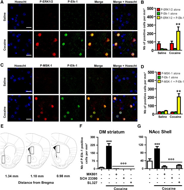
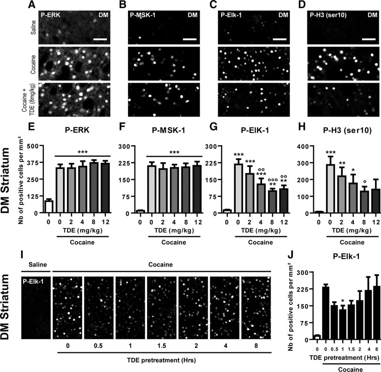
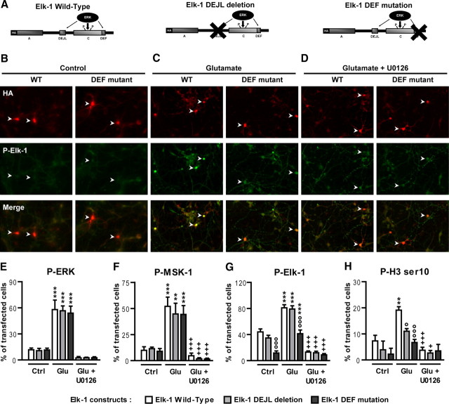
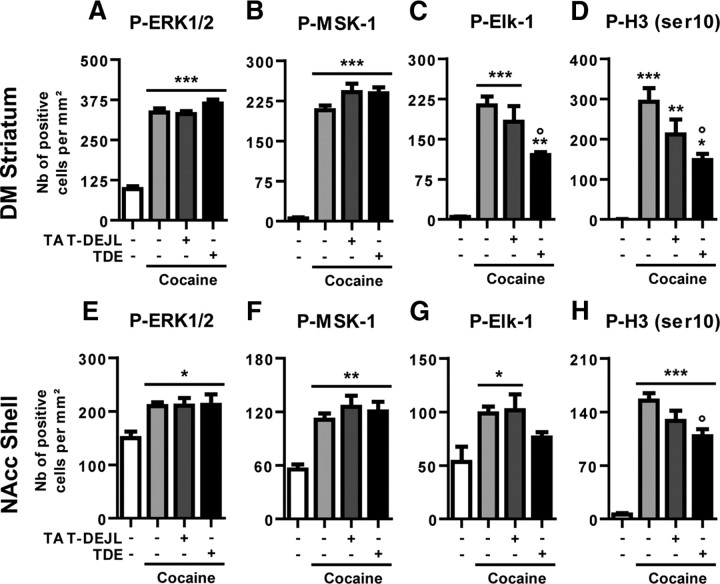
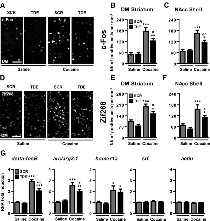
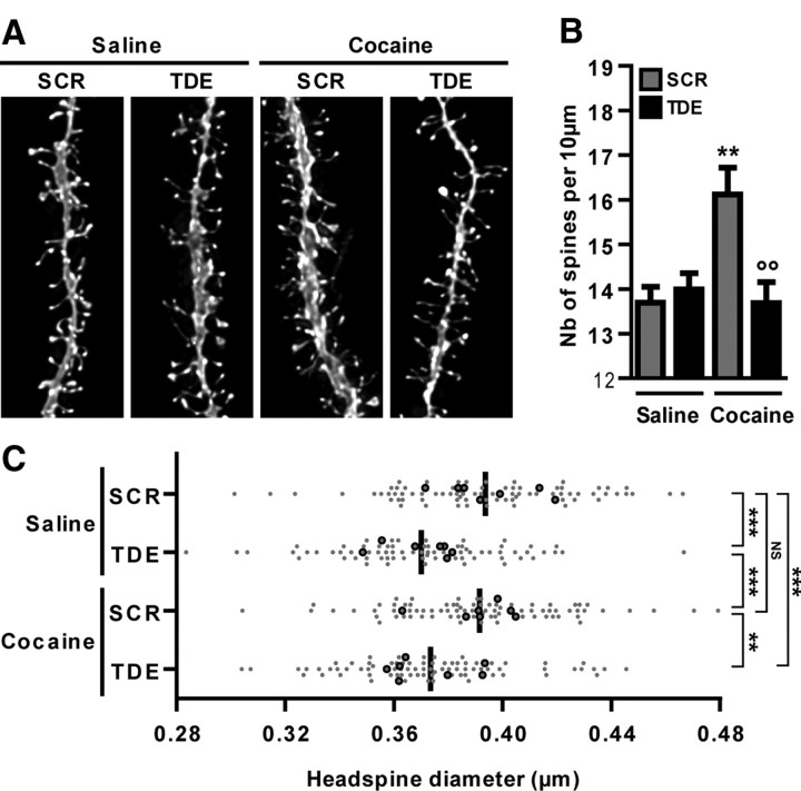
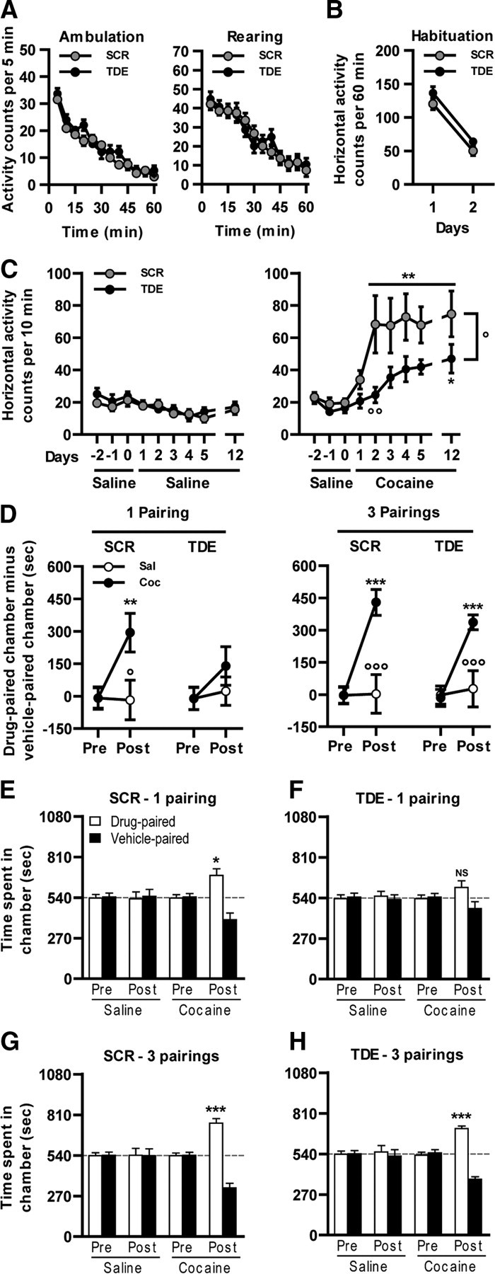
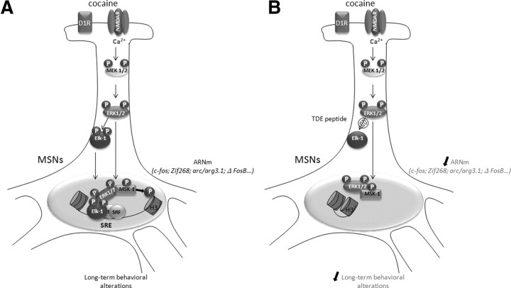
Activation of the extracellular signal-regulated kinase (ERK) signaling pathway in the striatum is crucial for molecular adaptations and long-term behavioral alterations induced by cocaine. In response to cocaine, ERK controls the phosphorylation levels of both mitogen and stress-activated protein kinase 1 (MSK-1), a nuclear kinase involved in histone H3 (Ser10) and cAMP response element binding protein phosphorylation, and Elk-1, a transcription factor involved in serum response element (SRE)-driven gene regulations. We recently characterized the phenotype of msk-1 knock-out mice in response to cocaine. Herein, we wanted to address the role of Elk-1 phosphorylation in cocaine-induced molecular, morphological, and behavioral responses. We used a cell-penetrating peptide, named TAT-DEF-Elk-1 (TDE), which corresponds to the DEF docking domain of Elk-1 toward ERK and inhibits Elk-1 phosphorylation induced by ERKs without modifying ERK or MSK-1 in vitro. The peptide was injected in vivo before cocaine administration in mice. Immunocytochemical, molecular, morphological, and behavioral studies were performed. The TDE inhibited Elk-1 and H3 (Ser10) phosphorylation induced by cocaine, sparing ERK and MSK-1 activation. Consequently, TDE altered cocaine-induced regulation of genes bearing SRE site(s) in their promoters, including c-fos, zif268, ΔFosB, and arc/arg3.1 (activity-regulated cytoskeleton-associated protein). In a chronic cocaine administration paradigm, TDE reversed cocaine-induced increase in dendritic spine density. Finally, the TDE delayed the establishment of cocaine-induced psychomotor sensitization and conditioned-place preference. We conclude that Elk-1 phosphorylation downstream from ERK is a key molecular event involved in long-term neuronal and behavioral adaptations to cocaine.


Figures

Figure 1.
Figure 1.
Patterning Elk-1 phosphorylation in response to cocaine in the striatum. A–D, Cocaine activates Elk-1 and MSK-1 in the same subset of striatal neurons. P-Elk-1 (A, C, green) was detected along with P-ERKs (A, red) or P-MSK-1 (C, red) by immunohistochemical detection on the same DM striatal section, 10 min after an acute treatment with saline (top rows) or cocaine (20 mg/kg, i.p.; bottom rows). The nuclei were labeled by Hoechst staining (blue). Images are single confocal sections. Scale bar, 25 μm. Note the strong colocalization of P-Elk-1 and P-ERK1/2 (A, merge) as well as P-Elk-1 and P-MSK-1 (C, merge) immunofluorescence occurring in the same subset of striatal cells (Merge + Hoescht). B–D, Quantifications of P-ERK1/2 and P-Elk-1 (B) or P-ERK1/2 and P-MSK-1 (D) immunoreactive cells. E, Schematic representation of striatal regions analyzed for quantifications in the DM and the NACC shell. Cocaine-induced P-Elk-1 immunoreactivity in the DM (F) or NAcc shell (G) is abolished by pharmacological inhibition of NMDA (MK801; 0.1 mg/kg) or DA D1 (SCH23390; 0.1 mg/kg) receptors or MEK inhibition (SL327; 50 mg/kg). The inhibitors were administered intraperitoneally 30 min before cocaine. B–D, Data (means ± SEM; n = 4 mice per group) were analyzed using unpaired t test for each marker. **p < 0.01, cocaine group versus saline group. F–G, Data (means ± SEM; n = 3–6 mice per group) were analyzed using one-way ANOVA (between subjects) for the DM (F(4,14) = 34.38, p < 0.001) and NAcc shell (F(4,14) = 18.81, p < 0.001), followed by post hoc comparisons (Newman–Keuls test). ***p < 0.001, cocaine group versus saline group; °°°p < 0.001, cocaine group versus cocaine groups pretreated with antagonists or MEK inhibitor.
Figure 2.
Figure 2.
Dose–response curves and kinetics of TDE effects on cocaine-induced phosphorylation of ERK, MSK-1, Elk-1, and H3 (Ser10). A–D, Immunohistochemical detection of cocaine-induced P-ERK1/2 (A), P-MSK-1 (B), P-Elk-1 (C), and P-H3 (Ser10) (D) was performed on striatal slices (illustrated here is the DM) 10 min after an acute treatment with saline or cocaine (20 mg/kg, i.p.) in mice. When indicated, the mice were pretreated with the TDE peptide (8 mg/kg), 60 min before cocaine treatment. Note the increased immunoreactivity for all markers in the cocaine group compared with saline control group. Note also the decreased P-Elk-1 and P-H3 (Ser10) immunoreactivities in the TDE-treated cocaine group when compared with cocaine alone. Scale bars, 50 μm. E–H, Quantifications of P-ERK1/2 (E), P-MSK-1 (F), P-Elk-1 (G), and P-H3 (Ser10) (H) immunoreactive cells in the DM, 10 min after an acute treatment with saline, cocaine alone (20 mg/kg), or cocaine and TDE (0–12 mg/kg) 60 min before cocaine. I, Illustration of the TDE effects on cocaine-induced phosphorylation of Elk-1. TDE (8 mg/kg) administered from 0.5 to 8 h before cocaine (20 mg/kg); mice were killed 10 min after cocaine administration. J, Quantification of P-Elk-1 immunoreactive cells in the DM in mice pretreated with TDE at indicated times (0–8 h) before a cocaine treatment occurring 10 min before being killed. Note the limited time window of TDE inhibitory effect between 30 and 120 min before cocaine administration. E–H, Data (means ± SEM; n = 8–16 mice per group) were analyzed using one-way ANOVA (between subjects) for P-ERK1/2 (F(5,62) = 22.29, p < 0.001), P-MSK-1 (F(5,62) = 31.76, p < 0.001), P-Elk-1 (F(5,62) = 17.05, p < 0.001), and P-H3 (Ser10) (F(5,62) = 6.665, p < 0.001), followed by post hoc comparisons (Newman–Keuls test). *p < 0.05, **p < 0.01, ***p < 0.001, cocaine groups versus saline group; °p < 0.05, °°p < 0.01, °°°p < 0.001, TDE pretreated cocaine groups versus saline pretreated cocaine group. J, Data (means ± SEM; n = 7–20 mice per group) were analyzed using one-way ANOVA (between subjects) (F(6,65) = 2.254, p < 0.05), followed by post hoc comparisons (Dunnett's multiple test). *p < 0.05, 0.5–8 h TDE pretreated groups versus immediately TDE pretreated group.
Figure 3.
Figure 3.
Mutations of Elk-1 DEF or DEJL domain alter glutamate-induced H3 (Ser10) phosphorylation but preserve MSK-1 activation in vitro. A, Schematic representation of the Elk-1 mutants encoded by the plasmids used for transfections. Striatal neurons were transfected at DIV6 with cDNAs encoding HA-tagged versions of WT Elk-1 (Elk-1 Wild-Type), Elk-1 carrying a deletion of the DEJL domain (Elk-1 DEJL deletion), or a mutation of the DEF domain (Elk-1 DEF mutation). Twenty-four hours later, neurons were incubated in the absence (Control) or presence of glutamate (Glutamate; 10 μm) for 20 min, with or without U0126 (10 μm) applied 30 min before and during glutamate (Glutamate + U0126) application. B–D, Representative pictures of striatal neurons transfected with the WT or HA-tagged DEF Elk-1 mutants treated or not with glutamate in the absence or presence of U0126. Transfected cells were detected with anti-HA antibody (red) or P-Elk-1 with a phospho-specific antibody (green). White arrows indicate transfected striatal neurons that are P-Elk-1 positive. E–H, Quantifications of P-ERK1/2 (E), P-MSK-1 (F), P-Elk-1 (G), and P-H3 (Ser10) (H) immunoreactive cells, in HA-transfected cells. Note the strong decrease in P-Elk-1 and P-H3 (Ser10) signals in cells transfected with the DEF mutant, whereas the DEJL mutant only affects P-H3 (Ser10). Note also the total inhibition of all markers in the case of a pretreatment with U0126. E–H, Data (means ± SEM; n = 3–4 independent experiments per group) were analyzed using two-way ANOVA: for P-ERK1/2, effect of treatment, F(2,18) = 95.28, p < 0.001; effect of mutation, F(2,18) = 0.059, NS; for P-MSK-1: effect of treatment, F(2,18) = 53.71, p < 0.001; effect of mutation, F(2,18) = 0.39, NS; for P-Elk-1: effect of treatment, F(2,27) = 120.4, p < 0.001; effect of mutation, F(2,27) = 29.54, p < 0.001; for P-H3 (Ser10): effect of treatment, F(2,18) = 19.05, p < 0.001; effect of mutation, F(2,18) = 7.36, p < 0.01, followed by post hoc comparisons (Bonferroni's test). **p < 0.01, ***p < 0.001, control (Ctrl) versus glutamate (Glu); +p < 0.05, +++p < 0.001, glutamate versus glutamate + U0126; °p < 0.05, °°°p < 0.001, wild-type Elk-1 versus Elk-1 construct (DEF mutation or DEJL deletion).
Figure 4.
Figure 4.
Comparative effects of TDE and TAT–DEJL–Elk-1 peptide on molecular responses to cocaine. Quantification of P-ERK1/2 (A, E), P-MSK-1 (B, F), P-Elk-1 (C, G), and P-H3 (Ser10) (D, H) immunoreactive cells in the DM (A–D) or in the NAcc shell (E–H), 10 min after an acute treatment with saline or cocaine (20 mg/kg). When indicated, the mice were pretreated with the TDE or the TAT–DEJL–Elk-1 (TAT–DEJL) (8 mg/kg, i.p.) 60 min before cocaine treatment. Note the increased immunoreactivity for all of the markers in the cocaine groups compared with saline control groups. Note also the decreased P-Elk-1 and P-H3 (Ser10) immunoreactivities in the cocaine group pretreated with TDE when compared with cocaine alone and the absence of effect of TAT–DEJL–Elk-1 on P-Elk-1 immunoreactivity. A–H, Data (means ± SEM; n = 3–7 mice per group) were analyzed using one-way ANOVA (between subjects) in the DM for P-ERK1/2 (F(3,14) = 91.30, p < 0.001), P-MSK-1 (F(3,14) = 103.3, p < 0.001), P-Elk-1 (F(3,14) = 25.63, p < 0.001), and P-H3 (Ser10) (F(3,14) = 15.99, p < 0.001) and in the NAcc shell for P-ERK1/2 (F(3,14) = 5.99, p < 0.01), P-MSK-1 (F(3,14) = 11.46, p < 0.001), P-Elk-1 (F(3,14) = 4.52, p < 0.05), and P-H3 (Ser10) (F(3,14) = 41.20, p < 0.001), followed by post hoc comparisons (Newman–Keuls test). *p < 0.05, **p < 0.01, ***p < 0.001, cocaine groups versus saline group; °p < 0.05, TDE pretreated cocaine group versus saline pretreated cocaine group.
Figure 5.
Figure 5.
Inhibition of Elk-1 phosphorylation alters SRE-driven gene regulation by cocaine in vivo. Immunohistochemical detections of c-Fos (A–C) and Zif268 (D–F) were performed in the DM and the NAcc shell 60 min after an acute treatment with saline or cocaine (10 mg/kg) in mice pretreated with TDE or SCR (8 mg/kg) 60 min before cocaine treatment. Note the increased immunoreactivity of c-Fos (A) and Zif268 (D) in mice pretreated with SCR before cocaine administration. Note also the decreased immunoreactivity in TDE pretreated cocaine groups. Scale bars, 50 μm. G, Real-time quantitative PCR was performed to evaluate levels of ΔfosB, arc/arg3.1, homer1a, srf, and actin mRNA 45 min after an acute cocaine injection in mice pretreated for 1 h with either TDE or SCR. B, C, E, F, Data (means ± SEM; n = 8 mice per group) were analyzed using one-way ANOVA (between subjects) in the DM for c-Fos (F(3,28) = 16.54, p < 0.001) and Zif268 (F(5,21) = 16.80, p < 0.001) and the NAcc shell for c-Fos (F(3,28) = 14.51, p < 0.001) and Zif268 (F(3,28) = 10.86, p < 0.001), followed by post hoc comparisons (Newman–Keuls test). *p < 0.05, **p < 0.01, ***p < 0.001, cocaine groups versus saline group; °p < 0.05, °°p < 0.01, TDE pretreated cocaine group versus SCR pretreated cocaine group. G, Data (means ± SEM; n = 5–6 mice per group) were analyzed using one-way ANOVA (between subjects) for ΔfosB (F(3,19) = 34.87, p < 0.001), arc/arg3.1 (F(3,19) = 16.64, p < 0.001), homer1a (F(3,19) = 5.349, p < 0.01), srf (F(3,19) = 0.581, NS), and actin (F(3,19) = 0.163, NS), followed by post hoc comparisons (Newman–Keuls test). *p < 0.05, **p < 0.01, ***p < 0.001, cocaine groups versus saline group; °p < 0.05, °°p < 0.01, TDE pretreated cocaine group versus SCR pretreated cocaine group.
Figure 6.
Figure 6.
Inhibition of Elk-1 phosphorylation inhibits cocaine-induced dendritic plasticity. A three-dimensional morphological analysis of DiI-labeled medium spiny neurons was performed in the NAcc shell of mice chronically treated after a chronic treatment with saline or cocaine (10 mg/kg; 5 injections over 3 d) in mice pretreated with TDE or SCR (8 mg/kg) 60 min before cocaine treatment. A, Representative pictures of DiI-stained dendritic sections in each experimental group. B, Quantifications of the number of dendritic spines detected per 10 μm of dendritic sections. Note that pretreatment with TDE totally abrogates cocaine-induced increase in dendritic spine density. C, Quantifications of head-spine diameters for each experimental group. Black dots represent the mean head-spine diameters per animal (n = 7 animals per group), and gray dots represent the median head-spine diameters per neuron (n = 10–14 neurons per animal). The black bar represents the mean head-spine diameters per group. Note the decreased head-spine diameters in chronically TDE pretreated groups independently of cocaine. B, Data (means ± SEM; n = 7 mice per group) were analyzed using two-way ANOVA: effect of pretreatment, F(1,24) = 5.443, p < 0.05; effect of treatment, F(1,24) = 5.342, p < 0.05, followed by post hoc comparisons (Bonferroni's test). **p < 0.01, cocaine groups versus saline group; °°p < 0.01, TDE pretreated cocaine group versus SCR pretreated cocaine group. C, Data (n = 72–79 neurons) were analyzed using Kruskal–Wallis test (p < 0.0001), followed by post hoc comparisons (Dunn's tests). **p < 0.01, ***p < 0.001; NS, not significant.
Figure 7.
Figure 7.
Inhibition of Elk-1 phosphorylation and cocaine-induced behavioral adaptations. Spontaneous locomotor activity: TDE compared with SCR injected at the dose of 8 mg/kg 60 min before the exposure to a novel environment (actimeter) does not alter either the spontaneous horizontal (ambulation) or vertical activity (rearing). B, Locomotor habituation. TDE compared with SCR injected at the dose of 8 mg/kg 60 min before the exposure to the same environment (actimeter) for 2 consecutive days does not alter the locomotor habituation. C, After 3 habituation days (days −2 to 0), mice were pretreated daily for 5 consecutive days, with either TDE or SCR at the dose of 8 mg/kg 60 min before saline (left) or cocaine (10 mg/kg; right) injection, and locomotor activity was measured. This procedure was also repeated on day 12 after a 7 d withdrawal. D, E, CPP induced by cocaine (n = 8 mice per group) was performed from mice pretreated during the conditioning phase of the protocol with TDE or its scramble version 1 h before cocaine administration in a one-pairing (D, left; E, F) or three-pairing (D, right; G, H) CPP paradigm. C, Data (means ± SEM; n = 14–15 mice per group) were analyzed using mixed-factor two-way ANOVA (repeated measure over time): saline: effect of time, F(8,224) = 7.53, p < 0.001; effect of pretreatment, F(1,224) = 0.35, NS; 10 mg/kg cocaine: effect of time, F(8,216) = 10.69, p < 0.001; effect of pretreatment, F(1,216) = 6.55, p < 0.05, followed by post hoc comparisons (Bonferroni's test). *p < 0.05, **p < 0.01, present day versus day 1; °p < 0.05, °°p < 0.01, TDE pretreated cocaine group versus SCR pretreated cocaine group. D, Data (means ± SEM; n = 8 mice per group) were analyzed using mixed-factor two-way ANOVA (repeated measure over conditioning session): one-pairing protocol SCR pretreatment: effect of conditioning, F(1,14) = 7.37, p < 0.05; effect of treatment, F(1,14) = 3.12, NS; one-pairing protocol TDE pretreatment: effect of conditioning, F(1,14) = 2.51, NS; effect of treatment, F(1,14) = 0.62, NS; three-pairing protocol SCR pretreatment: effect of conditioning, F(1,14) = 32.68, p < 0.001; effect of treatment, F(1,14) = 7.73, p < 0.05; three-pairing protocol TDE pretreatment: effect of conditioning, F(1,14) = 21.0, p < 0.001; effect of treatment, F(1,14) = 5.35, p < 0.05, followed by post hoc comparisons (Bonferroni's test). **p < 0.01, ***p < 0.001, posttest versus pretest; °p < 0.05, °°°p < 0.001, saline versus cocaine. E–H, Data (means ± SEM; n = 8 mice per group) were analyzed using paired t test for all of the groups before (Pre) and after (Post) conditioning. *p < 0.05, **p < 0.01, ***p < 0.001, drug-paired chamber versus vehicle-paired chamber. Dashed lines represent equal exploration of both chambers.
Figure 8.
Figure 8.
Schematic representation of molecular events driven by Elk-1 phosphorylation in response to cocaine. A, During cocaine administration, combined activation of D1R and NMDAR drives ERK activation via calcium entry (Pascoli et al., 2011). This leads to the phosphorylation and nuclear translocation of Elk-1 along with ERKs. Within the nucleus, Elk-1 binds to the SRE sites of IEGs (including c-fos, zif268, and arc/arg3.1) and recruits ERK1/2 and MSK-1 at the proximity of the nucleosome. In turn, MSK-1 can phosphorylate histone H3 on Ser10 residue, allowing nucleosome repositioning and transcription. These molecular events are critically involved in MSN spine plasticity and long-term behavioral alterations in response to cocaine. B, In the presence of the TDE peptide, Elk-1 phosphorylation and nuclear translocation is impeded. Cocaine-induced ERK and MSK-1 phosphorylation persist, but MSK-1 is not recruited to the nucleosome. SRE-driven gene regulation is inhibited, along with spine plasticity and long-term behavioral responses.

References

    1. Bertran-Gonzalez J, Bosch C, Maroteaux M, Matamales M, Hervé D, Valjent E, Girault JA. Opposing patterns of signaling activation in dopamine D1 and D2 receptor-expressing striatal neurons in response to cocaine and haloperidol. J Neurosci. 2008;28:5671–5685. - PMC - PubMed
    1. Bramham CR, Worley PF, Moore MJ, Guzowski JF. The immediate early gene arc/arg3.1: regulation, mechanisms, and function. J Neurosci. 2008;28:11760–11767. - PMC - PubMed
    1. Brami-Cherrier K, Valjent E, Garcia M, Pagès C, Hipskind RA, Caboche J. Dopamine induces a PI3-kinase-independent activation of Akt in striatal neurons: a new route to cAMP response element-binding protein phosphorylation. J Neurosci. 2002;22:8911–8921. - PMC - PubMed
    1. Brami-Cherrier K, Valjent E, Hervé D, Darragh J, Corvol JC, Pages C, Arthur SJ, Girault JA, Caboche J. Parsing molecular and behavioral effects of cocaine in mitogen- and stress-activated protein kinase-1-deficient mice. J Neurosci. 2005;25:11444–11454. [Erratum (2006) 26:table of contents; Simon, Arthur J corrected to Arthur, Simon J] - PMC - PubMed
    1. Brami-Cherrier K, Roze E, Girault JA, Betuing S, Caboche J. Role of the ERK/MSK1 signalling pathway in chromatin remodelling and brain responses to drugs of abuse. J Neurochem. 2009;108:1323–1335. - PubMed

Publication types