Comparative Study
. 2011 Oct 5;31(40):14436-49.
doi: 10.1523/JNEUROSCI.3836-11.2011.

Chronic stress exacerbates tau pathology, neurodegeneration, and cognitive performance through a corticotropin-releasing factor receptor-dependent mechanism in a transgenic mouse model of tauopathy

Affiliations
Comparative Study

Chronic stress exacerbates tau pathology, neurodegeneration, and cognitive performance through a corticotropin-releasing factor receptor-dependent mechanism in a transgenic mouse model of tauopathy

Jenna C Carroll et al. J Neurosci. .

Abstract

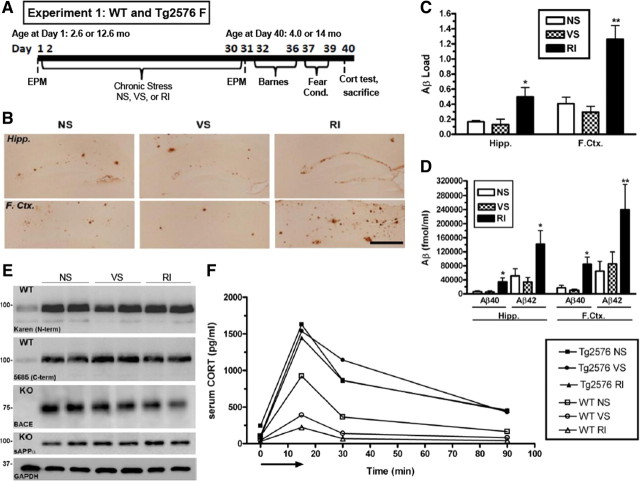
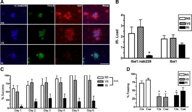
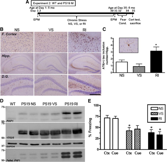
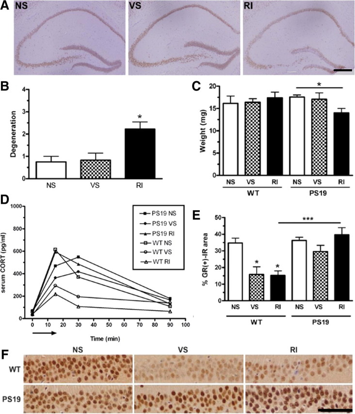
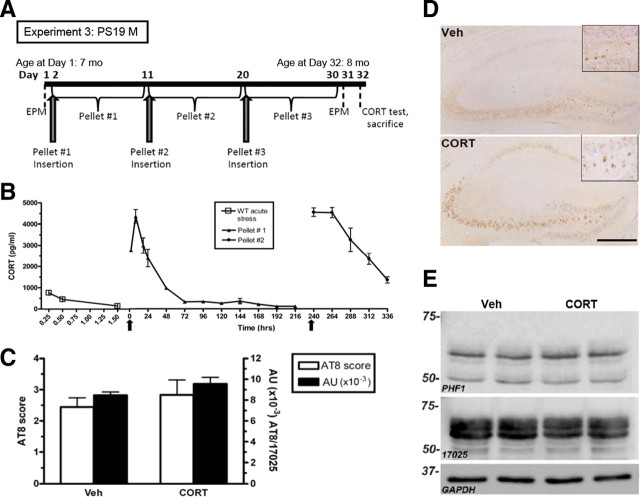
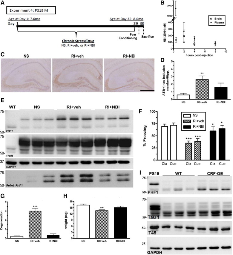
Because overactivation of the hypothalamic-pituitary-adrenal (HPA) axis occurs in Alzheimer's disease (AD), dysregulation of stress neuromediators may play a mechanistic role in the pathophysiology of AD. However, the effects of stress on tau phosphorylation are poorly understood, and the relationship between corticosterone and corticotropin-releasing factor (CRF) on both β-amyloid (Aβ) and tau pathology remain unclear. Therefore, we first established a model of chronic stress, which exacerbates Aβ accumulation in Tg2576 mice and then extended this stress paradigm to a tau transgenic mouse model with the P301S mutation (PS19) that displays tau hyperphosphorylation, insoluble tau inclusions and neurodegeneration. We show for the first time that both Tg2576 and PS19 mice demonstrate a heightened HPA stress profile in the unstressed state. In Tg2576 mice, 1 month of restraint/isolation (RI) stress increased Aβ levels, suppressed microglial activation, and worsened spatial and fear memory compared with nonstressed mice. In PS19 mice, RI stress promoted tau hyperphosphorylation, insoluble tau aggregation, neurodegeneration, and fear-memory impairments. These effects were not mimicked by chronic corticosterone administration but were prevented by pre-stress administration of a CRF receptor type 1 (CRF(1)) antagonist. The role for a CRF(1)-dependent mechanism was further supported by the finding that mice overexpressing CRF had increased hyperphosphorylated tau compared with wild-type littermates. Together, these results implicate HPA dysregulation in AD neuropathogenesis and suggest that prolonged stress may increase Aβ and tau hyperphosphorylation. These studies also implicate CRF in AD pathophysiology and suggest that pharmacological manipulation of this neuropeptide may be a potential therapeutic strategy for AD.


Conflict of interest statement

The authors declare no conflicts of interest.

Figures

Figure 1.
Figure 1.
Chronic RI stress exacerbates Aβ levels in stress-sensitive Tg2576 mice. A, Experimental design of experiment 1; WT and Tg2576 female mice (n = 10 per group) were exposed to NS, VS, or RI stress for 1 month. B, Representative images in the hippocampus (Hipp.) and frontal cortex (F.Ctx.) show increased Aβ-IR in RI mice, quantified in C (F(3,18) = 5.61, p = 0.014; and F(3,24) = 15.64, p < 0.0001). Data show mean ± SEM Aβ load. D, By ELISA, insoluble Aβ40 was significantly increased by RI in the hippocampus (F(5,28) = 4.47, p = 0.005) and frontal cortex (F(5,28) = 2.08, p = 0.11), as was Aβ42 in the hippocampus (F(2,21) = 6.51, p = 0.007) and frontal cortex (F(2,15) = 12.4, p = 0.001). Data show mean ± SEM Aβ levels (n = 8 per group). E, Hippocampal protein levels of APP, sAPPα, and BACE were measured with either WT or BACE KO mouse hippocampal homogenate (n = 2 per group) as a negative control. In 14-month-old Tg2576 mice, neither stress altered hippocampal levels of 5685, Karen, BACE, or sAPP. F, CORT levels were plotted over time after an acute 15 min restraint stress (arrow) in 4-month-old WT and Tg2576 mice. In WT mice, both VS (F(5,28) = 4.9, p = 0.05) and RI stress (F(2,20) = 1.3, p = 0.31) decreased the area under the curve compared with NS. However, CORT levels were elevated significantly in all conditions of Tg2576 mice, regardless of stress treatment. Data show mean (n = 10 per group). *p < 0.05, **p < 0.0 1 from NS. Scale bar, 0.5 mm.
Figure 2.
Figure 2.
Chronic stress impairs spatial and fear-related learning but does not correlate with activated microglia-associated Aβ plaques in Tg2576 mice. Tg2576 mice were assessed for activated microglia associated with Aβ plaques. A, Representative images show that, regardless of stress condition, all mice display Nab228-IR Aβ plaques (blue) that are Thioflavin-S+ (green) in the frontal cortex. However, RI mice showed significantly less Iba1-IR activated microglia (red) around Aβ plaques without a decrease in overall Iba1-IR. B, This effect is quantified by nonfluorescent immunoreactivity load ratios (mean ± SEM) of adjacent sections (F(2,21) = 6.51, p = 0.007; and F(2,21) = 6.51, p = 0.007; n = 8 per group). Fourteen-month-old Tg2576 mice (n = 10 per group) were also tested on the Barnes maze (C) and fear conditioning (D). RI but not VS mice demonstrated significantly worse percentage success (F(3,35) = 12.8, p < 0.0001) compared with NS mice. In both context (Ctx) (F(2,20) = 6.22, p = 0.009) and cued (Cue) (F(2,21) = 5.98, p = 0.01) fear conditioning, both VS and RI stressed Tg2576 mice froze significantly less than NS mice. Data show mean ± SEM. *p < 0.05, ***p < 0.001 from NS. Scale bar, 100 μm.
Figure 3.
Figure 3.
Chronic RI stress exacerbates hyperphosphorylated tau, aggregated tau inclusions, and fear-related memory in PS19 mice. A, Experimental design of experiment 2; WT and PS19 male mice (n = 8 per group). B, Representative images demonstrate that, in the frontal cortex (F. Cortex) and the hippocampus (Hipp.), specifically the dentate gyrus (D.G.), chronic RI increased AT8-IR compared with NS. C, The number of AT8-IR tau inclusions (inset) in the hippocampus was counted and showed a significant increase in RI compared with NS mice (F(2,23) = 14.42, p < 0.0001). Data show mean ± SEM number of tau inclusions. D, Tau was also evaluated by Western blot using both PHF1 and 17025 antibodies and probed for GAPDH as a loading control. PS19 RI stressed mice displayed a significant increase in soluble PHF1 (F(2,5) = 8.21, p = 0.0024) and insoluble (F(2,5) = 1.96, p = 0.009) but not 17025 expressions in the hippocampus. E, PS19 RI mice showed significant impairment compared with NS mice in both context (Ctx) (F(5,54) = 3.88, p = 0.0048) and cued (Cue) fear conditioning (F(5,54) = 4.19, p = 0.003). *p < 0.05, from NS of same genotype. Scale bar (in B): 0.5 mm for cortex and hippocampus; 200 mm for dentate gyrus.
Figure 4.
Figure 4.
Chronic RI stress induces neurodegeneration, altered GR expression, and altered corticosterone response in PS19 mice. PS19 mice were evaluated for hippocampal neurodegeneration. A, Representative pictomicrographs of NeuN-IR show that PS19 RI mice displayed significantly higher degeneration as quantified in B (F(2,18) = 6.89, p = 0.007). C, Hemi-hippocampal weight assessment also revealed that PS19 (but not WT) RI mice displayed lower hippocampal weight (F(2,21) = 3.62, p = 0.04) compared with VS or NS mice. Data show mean ± SEM (n = 8 per group). *p < 0.05 from NS. Scale bar, 0.5 mm. D, CORT levels were plotted over time after an acute 15 min restraint stress (arrow). In WT mice, both VS (F(2,18) = 24.7, p < 0.001) and RI (F(2,18) = 24.7, p < 0.001) stress decreased the area under the curve compared with NS. However, CORT levels were elevated significantly in all conditions of PS19 mice, regardless of stress treatment. Data show mean (n = 10 per group). F, These mice were also assessed for GR-IR in the CA1. Representative pictomicrographs of both WT and PS19 mice under different stress conditions show that VS and RI significantly reduced GR levels in WT but not PS19 mice. E, This effect was quantified to demonstrate a significant group effect (F(5,25) = 11.6, p < 0.0001). Data show mean ± SEM GR-IR load. *p < 0.05, and ***p < 0.001. Scale bar, 50 μm.
Figure 5.
Figure 5.
CORT treatment does not mimic chronic stress-induced tau hyperphosphorylation in PS19 mice. A, Experimental design of experiment 3; PS19 male mice were not stressed but were administered CORT pellets (n = 8 per group). B, First, a small pilot (n = 3 per group) of adult WT male mice were implanted with CORT pellets (arrows) to assess the plasma hormone levels achieved by these pellets. A high spike in CORT within the first 72 h was significantly higher (F(2,7) = 2.25, p < 0.0001) than after an acute stress from experiment 1 (data also shown in Fig. 4A). Furthermore, neither representative images of AT8-IR in the hippocampus (C) nor IR quantification using a semiquantitative rating scale (0–5) (D) showed a treatment difference. AU, Arbitrary units; Veh, vehicle. Data show mean ± SEM AT8 score. E, A representative Western blot probed for both PHF1, 17025, and GAPDH as a loading control also demonstrate no difference after CORT treatment in the hippocampus. Scale bar, 0.5 mm.
Figure 6.
Figure 6.
CRF1 mediates stress-induced pathological tau inclusions, neurodegeneration, and fear-associated learning. A, Experimental design of experiment 4; male PS19 mice were exposed to NS or RI stress with pre-stress CRF1 antagonist (NBI) or vehicle subcutaneous injections (n = 8 per group). B, A small pilot study using adult WT female mice (n = 3 per group) was conducted to measure brain and plasma levels of NBI after a 10 mg/kg subcutaneous injection. C, Representative pictomicrographs demonstrate that vehicle-treated RI mice displayed significantly higher AT8-IR tau inclusions in the hippocampus, quantified in D (F(2,25) = 7.15, p = 0.05), as well as soluble and insoluble PHF1, but not 17025 levels by Western blot (E) that was blocked by a pre-stress injection of NBI. F, Vehicle-treated RI mice also displayed fear-related learning impairments in both context (Ctx) (F(2,28) = 12.4, p < 0.05) and cued (Cue) (F(2,28) = 9.3, p < 0.05) fear conditioning that was significantly prevented by NBI. Data show mean ± SEM percentage freezing. G, Compared with NS, vehicle-treated mice had significantly lower hippocampal weight (F(2,44) = 6.44, p = 0.004) and significantly increased NeuN+-IR degeneration scores (F(2,28) = 6.19, p < 0.001) (H), whereas NBI treatment significantly prevented both effects. I, Last, phosphorylated tau was quantified in the hippocampus of 10-month-old CRF-OE mice and WT littermate controls. A substantial elevation was observed in PHF1 levels in CRF-OE compared with WT littermates without a change in Tau 1 and T49 level. Data show mean ± SEM PHF1 score. Scale bar, 0.5 mm. **p < 0.01, ***p < 0.001 from NS. *p < 0.05 from RI + vehicle.

Comment in

References

    1. Alfarez DN, Joëls M, Krugers HJ. Chronic unpredictable stress impairs long-term potentiation in rat hippocampal CA1 area and dentate gyrus in vitro. Eur J Neurosci. 2003;17:1928–1934. - PubMed
    1. Arendt T, Stieler J, Strijkstra AM, Hut RA, Rüdiger J, Van der Zee EA, Harkany T, Holzer M, Härtig W. Reversible paired helical filament-like phosphorylation of tau is an adaptive process associated with neuronal plasticity in hibernating animals. J Neurosci. 2003;23:6972–6981. - PMC - PubMed
    1. Avila J, Wandosell F, Hernández F. Role of glycogen synthase kinase-3 in Alzheimer's disease pathogenesis and glycogen synthase kinase-3 inhibitors. Expert Rev Neurother. 2010;10:703–710. - PubMed
    1. Aznar S, Knudsen GM. Depression and Alzheimer's disease: is stress the initiating factor in a common neuropathological cascade? J Alzheimers Dis. 2011;23:177–193. - PubMed
    1. Bachman DL, Wolf PA, Linn R, Knoefel JE, Cobb J, Belanger A, D'Agostino RB, White LR. Prevalence of dementia and probable senile dementia of the Alzheimer type in the Framingham Study. Neurology. 1992;42:115–119. - PubMed

Publication types

MeSH terms

Substances