. 2012 Feb 14;86(2):37.
doi: 10.1095/biolreprod.111.095208. Print 2012 Feb.

Temporal differences in granulosa cell specification in the ovary reflect distinct follicle fates in mice

Affiliations

Temporal differences in granulosa cell specification in the ovary reflect distinct follicle fates in mice

Lindsey Mork et al. Biol Reprod. .

Abstract

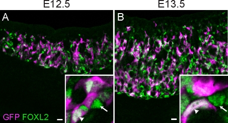
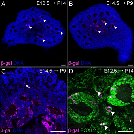
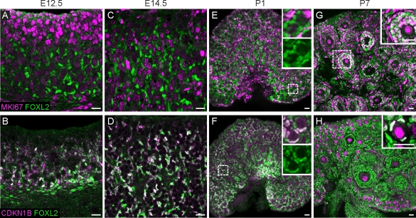
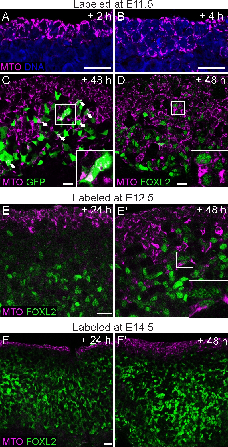
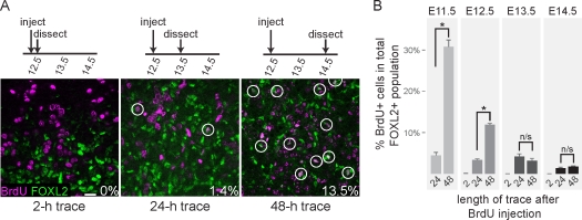
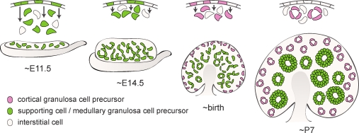
The embryonic origins of ovarian granulosa cells have been a subject of debate for decades. By tamoxifen-induced lineage tracing of Foxl2-expressing cells, we show that descendants of the bipotential supporting cell precursors in the early gonad contribute granulosa cells to a specific population of follicles in the medulla of the ovary that begin to grow immediately after birth. These precursor cells arise from the proliferative ovarian surface epithelium and enter mitotic arrest prior to upregulating Foxl2. Granulosa cells that populate the cortical primordial follicles activated in adult life derive from the surface epithelium perinatally, and enter mitotic arrest at that stage. Ingression from the surface epithelium dropped to undetectable levels by Postnatal Day 7, when most surviving oocytes were individually encapsulated by granulosa cells. These findings add complexity to the standard model of sex determination in which the Sertoli and granulosa cells of the adult testis and ovary directly stem from the supporting cell precursors of the bipotential gonad.


Figures

FIG. 1.
FIG. 1.
FOXL2 is expressed by supporting cell precursors that are competent to express Sry. Ovaries from E12.5 (A) and E13.5 (B) XX Sry-EGFP transgenic embryos immunostained with antibodies against FOXL2 (green, nuclear) and EGFP (magenta, nuclear and cytoplasmic) show clear overlap (white) between the two markers (arrowheads in insets), although some FOXL2-positive cells were not EGFP positive (arrows in insets). Original magnification ×20; bars = 20 μm.
FIG. 2.
FIG. 2.
Foxl2-expressing cells in the fetal ovary give rise to granulosa cells in medullary follicles. AC) Foxl2-GCE; R26R mice were exposed to tamoxifen at E12.5 and E14.5, dissected at P14 (A) or P9 (B and C), and stained with an antibody against β-galactosidase (magenta). Positive staining was nearly exclusive to granulosa cells in the large follicles in the medulla of the ovary. Arrow in C indicates a very rare lineage-traced granulosa cell in a primordial follicle. D) High-magnification image of a P14 Foxl2-GCE; R26R ovary from a mouse exposed to tamoxifen at E12.5, stained with antibodies against β-galactosidase (magenta) and FOXL2 (green). Lineage-labeled cells were observed in large secondary follicles as well as smaller primary follicles, but not primordial follicles. Arrowheads indicate growing follicles containing lineage-labeled cells that were sectioned through the edge rather than the middle of the follicle. Nuclei (blue) were stained with syto13. Original magnification ×10 (A and B) or ×40 (C and D); bars = 50 μm.
FIG. 3.
FIG. 3.
FOXL2-positive cells are arrested throughout embryonic development. Cells expressing FOXL2 (green) were negative for MKI67 (magenta; A and C) and positive for CDKN1B (magenta; B and D) at E12.5 and E14.5. At birth (E and F), the FOXL2-positive cells in close proximity to developing oocytes remained arrested, although cells more distant from oocytes were positive for MKI67 and negative for CDKN1B. Insets in E and F show FOXL2 expression in primordial follicles and surrounding interstitial cells, with or without MKI67 (E) or CDKN1B (F) overlay. By P7 (G and H), a subset of follicles had progressed into primary and secondary stages. FOXL2-positive cells in these activated follicles were MKI67-positive (inset in G) and CDKN1B-negative, although granulosa cells in primordial follicles in the cortical region of the gonad remained arrested (inset in H). Oocytes upregulated CDKN1B shortly after birth (F and H), as previously reported [32]. White color indicates overlap between the markers. AD are images of whole-mount immunostained gonads taken at 40×. EH are images of cryosectioned gonads (original magnification ×20). Bars = 20 μm.
FIG. 4.
FIG. 4.
The surface epithelium is a source of new Sry-EGFP- and Foxl2-expressing cells in the fetal ovary. The ovarian surface was labeled with the cytoplasmic MitoTracker dye (MTO; magenta) at various stages of ovary development, and the samples were cultured for 2–72 h. A) Gonads from E11.5 embryos fixed after 2 h show that the dye is confined to the outermost cell layers. B) After 6 h, cell divisions in the coelomic epithelium generate labeled cells that move deeper inside the gonad. C) Ovary from an Sry-EGFP transgenic embryo labeled with MitoTracker at E11.5. Many EGFP-positive cells contained the label after 48 h of culture (overlap is white; arrowheads and inset). D) Similarly, in an E11.5 wild-type ovary cultured for 48 h, MitoTracker-labeled cells had ingressed to very deep layers, and many began to present nuclear FOXL2 staining (green; inset). E and E′) Gonads labeled at E12.5 displayed fewer ingressing cells that colabeled with FOXL2 after 24–48 h. F and F′) In samples labeled at E14.5, no ingression was observed after 24 h. Multiple cell layers were labeled after 48 h, but few ingressing cells were positive for FOXL2. Original magnification ×40 (AE) or ×20 (F); bars = 20 μm.
FIG. 5.
FIG. 5.
Cycling progenitor cells give rise to new Foxl2-expressing cells. A) Example of BrdU lineage tracing and data collection. In this series, pregnant females were injected with BrdU at E12.5, chased with thymidine 2 h later, and dissected after the indicated trace durations. Gonads were stained with antibodies against BrdU (magenta) and FOXL2 (green); white color indicates overlap between the markers. BrdU/FOXL2 double-positive cells (circles) were counted relative to the total number of FOXL2-positive cells and expressed as a percentage. Original magnification ×40; bar = 20 μm. B) Quantification of BrdU results. Embryonic stages listed above the bars indicate when BrdU was injected (n = 4–12 images per time point, from two to four independent gonad samples). Error bars indicate SEM. Embryos pulsed at E11.5 or E12.5 showed significantly higher proportions of BrdU/FOXL2 double-positive cells in 48-h traces relative to 24-h traces, but those injected at E13.5–E14.5 did not, suggesting that the number of new cells entering the population was proportionally much lower at later stages. *P < 0.001; n/s, not significant.
FIG. 6.
FIG. 6.
New granulosa cells arise in the ovarian cortex after birth. A and B) Ovaries of P7 pups exposed to BrdU at P1 (A) or P4 (B) and stained with antibodies against BrdU (magenta) and αlaminin (green) show BrdU-positive granulosa cells inside primordial follicles (arrows, insets). As expected, granulosa cells in actively dividing medullary follicles were heavily labeled in samples pulsed at P4, but not in samples pulsed at P1. We speculate that the label was titrated out during the week-long chase in the latter case. C and D) Somatic cells in and near the surface epithelium remain proliferative after birth. Ovaries from P1 mice were injected with BrdU 2 h prior to dissection and stained with antibodies against BrdU (magenta) and CDKN1B (C) or FOXL2 (D) (green). Arrowheads point to clusters of proliferative (CDKN1B-negative, BrdU-positive) FOXL2-negative somatic cells under the surface epithelium. Nuclei (blue) were stained with syto13. Original magnification ×20 (A and B) or ×40 (C and D); bars = 20 μm.
FIG. 7.
FIG. 7.
The ovarian surface epithelium gives rise to new granulosa cells during the follicle assembly period. A) Ovaries from P1, P3, and P7 pups were labeled with the cytoplasmic MitoTracker dye (MTO, magenta), cultured for 24–96 h, then fixed and stained with an antibody against FOXL2 (green, nuclear). Only the surface epithelium was labeled after 24 h, but obvious ingression of labeled cells was observed after 72–96 h in cultures started at P1 or P3, but not P7. Arrowheads point to FOXL2-positive cells labeled with MitoTracker. B) A few squamous MitoTracker-labeled FOXL2-positive cells (arrowheads) were incorporated into primordial follicles after 96 h in ovaries placed into culture at P3. Nuclei (blue) were stained with syto13. Original magnification ×20 (A) or ×40 (B); bars = 20 μm.
FIG. 8.
FIG. 8.
Origins of ovarian granulosa cells. Divisions in the surface epithelium of the bipotential gonad (E11.5) produce both interstitial cells (beige) and supporting cell precursors (green) that differentiate as Sertoli cells in the presence of a Y chromosome; in XX individuals, these cells move deep into the ovary and give rise to the granulosa cells in the medullary follicles activated immediately after their assembly at birth (green). As the ovary differentiates (∼E14.5), further divisions in the surface epithelium contribute more cells to the granulosa population, although they remain closer to the ovarian surface. The granulosa cells that populate the primordial follicles in the cortex (pink), which are activated in adult life, arise from the surface epithelium around birth. By P7, follicle assembly is mostly complete, and surface cells no longer move into the ovary.

Comment in

References

    1. Hirshfield AN. Development of follicles in the mammalian ovary. Int Rev Cytol 1991; 124: 43 101 - PubMed
    1. McLaren A. Development of the mammalian gonad: the fate of the supporting cell lineage. Bioessays 1991; 13: 151 156 - PubMed
    1. Albrecht KH, Eicher EM. Evidence that Sry is expressed in pre-Sertoli cells and Sertoli and granulosa cells have a common precursor. Dev Biol 2001; 240: 92 107 - PubMed
    1. Auersperg N, Wong AS, Choi KC, Kang SK, Leung PC. Ovarian surface epithelium: biology, endocrinology, and pathology. Endocr Rev 2001; 22: 255 288 - PubMed
    1. Liu CF, Liu C, Yao HH. Building pathways for ovary organogenesis in the mouse embryo. Curr Top Dev Biol 2010; 90: 263 290 - PMC - PubMed

Publication types

Substances