. 2011 Dec;85(24):12939-49.
doi: 10.1128/JVI.05177-11. Epub 2011 Oct 5.

Cellular poly(c) binding proteins 1 and 2 interact with porcine reproductive and respiratory syndrome virus nonstructural protein 1β and support viral replication

Affiliations

Cellular poly(c) binding proteins 1 and 2 interact with porcine reproductive and respiratory syndrome virus nonstructural protein 1β and support viral replication

Lalit K Beura et al. J Virol. 2011 Dec.

Abstract

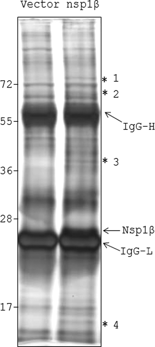
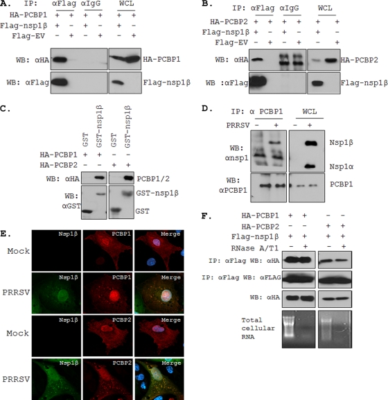
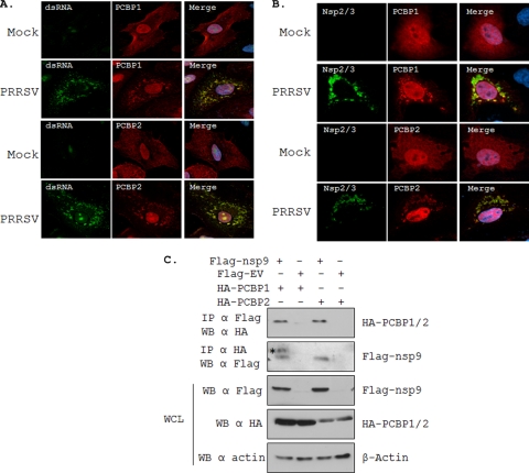
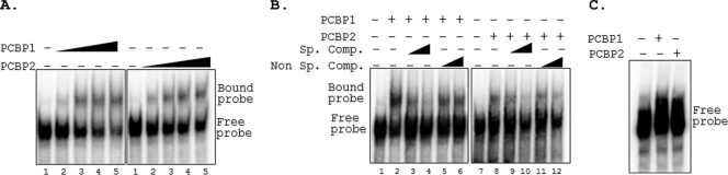
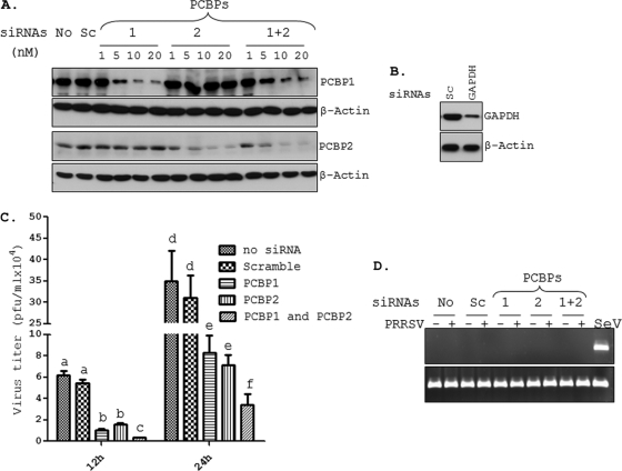
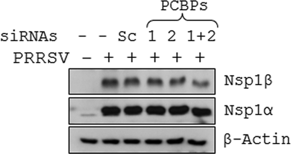
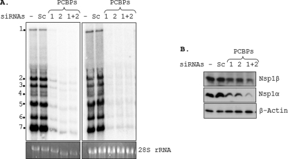
Porcine reproductive and respiratory syndrome virus (PRRSV) infection of swine results in substantial economic losses to the swine industry worldwide. Identification of cellular factors involved in PRRSV life cycle not only will enable a better understanding of virus biology but also has the potential for the development of antiviral therapeutics. The PRRSV nonstructural protein 1 (nsp1) has been shown to be involved in at least two important functions in the infected hosts: (i) mediation of viral subgenomic (sg) mRNA transcription and (ii) suppression of the host's innate immune response mechanisms. To further our understanding of the role of the viral nsp1 in these processes, using nsp1β, a proteolytically processed functional product of nsp1 as bait, we have identified the cellular poly(C)-binding proteins 1 and 2 (PCBP1 and PCBP2) as two of its interaction partners. The interactions of PCBP1 and PCBP2 with nsp1β were confirmed both by coimmunoprecipitation in infected cells and/or in plasmid-transfected cells and also by in vitro binding assays. During PRRSV infection of MARC-145 cells, the cytoplasmic PCBP1 and PCBP2 partially colocalize to the viral replication-transcription complexes. Furthermore, recombinant purified PCBP1 and PCBP2 were found to bind the viral 5' untranslated region (5'UTR). Small interfering RNA (siRNA)-mediated silencing of PCBP1 and PCBP2 in cells resulted in significantly reduced PRRSV genome replication and transcription without adverse effect on initial polyprotein synthesis. Overall, the results presented here point toward an important role for PCBP1 and PCBP2 in regulating PRRSV RNA synthesis.


Figures

Fig. 1.
Fig. 1.
Identification of PRRSV nsp1β-interacting proteins by IP-MS. Flag-EV (Vector)- and Flag-nsp1β (nsp1β)-overexpressing 293T cells were used for immunoprecipitation using anti-Flag agarose resins. Eluted proteins were resolved by 12% SDS-PAGE and subjected to silver staining. The prominent bands at ∼55 kDa and ∼25 kDa are IgG heavy (IgG-H) and IgG light (IgG-L) chains, respectively. The nsp1β band is observed just above the IgG-L band in the nsp1β lane only. The four bands specific to the nsp1β lane are indicated by asterisks (*). The relative mobility of molecular mass markers (in kDa) is shown on the left.
Fig. 2.
Fig. 2.
Interaction of PCBP1 and PCBP2 with PRRSV nsp1β. Co-IP of HA-PCBP1 (A) and HA-PCBP2 (B) with Flag-nsp1β. Indicated plasmids (shown on top) were transfected into 293T cells, and at 48 hpt, whole-cell lysates (WCL) were immunoprecipitated with anti-Flag or anti-IgG. After SDS-PAGE separation, proteins were detected by immunoblotting with antibodies indicated on the left. A 5% aliquot of WCL was also probed similarly to confirm the expression of the proteins. The identities of various protein bands are indicated on the right. (C) GST pulldown assay. Glutathione beads conjugated to GST or GST-nsp1β fusion protein were incubated with recombinant PCBP1 (left) or PCBP2 (right). After washing, proteins were eluted from the beads and SDS-PAGE was performed. The presence of PCBP1 and PCBP2 was detected by anti-HA antibody. GST and GST-nsp1β expression was confirmed by use of anti-GST antibody. (D) Co-IP of PRRSV nsp1β with endogenous PCBP1. PRRSV-infected (+) or mock-infected (−) MARC-145 cells were used for IP with anti-PCBP1 antibody and immunoblotted with antibodies shown on the left. The identities of various bands are shown on the right. (E) Colocalization of nsp1β with HA-PCBP1 and HA-PCBP2 by confocal microscopy. MARC-145 cells were mock infected or infected with PRRSV (the VR-2332 strain was used here, since the nsp1β monoclonal antibody is against this virus) for 6 h before being transfected with the PCBP protein-expressing plasmids. Cells were fixed 18 hpt and subjected to indirect immunofluorescence to detect nsp1β (green) and HA-PCBP1 or PCBP2 (red) by using mouse anti-nsp1β and rabbit anti-HA antibodies. Position of nucleus is indicated by DAPI (4′,6-diamidino-2-phenylindole) (blue) by staining in the merge image (far right). (F) Effect of RNase treatment on PCBP1/2-nsp1β interaction. Extracts of 293T cells overexpressing the different proteins were treated or untreated with RNase A and T1 for 1 h before IP and immunoblotted with antibodies shown on the left. Ethidium bromide-stained agarose gels show total RNA present with or without RNase treatment. WB, Western blotting.
Fig. 3.
Fig. 3.
Colocalization of PCBP1 and PCBP2 with dsRNA and nsp's in PRRSV-infected cells. (A) MARC-145 cells were mock infected or infected with PRRSV (strain VR-2332) for 6 h before being transfected with the PCBP protein-expressing plasmids. Cells were fixed 18 hpt and were subjected to indirect immunofluorescence to detect dsRNA (green) and HA-PCBP1 and HA-PCBP2 (red) by using mouse anti-dsRNA (J2) and rabbit anti-HA antibodies, respectively. (B) MARC-145 cells were treated as described above, and indirect immunofluorescence was performed to detect PCBP1 or PCBP2 (red) and PRRSV nsp2/3 (green) by using mouse anti-HA antibody and rabbit anti-nsp2/3 antibody, respectively. Nuclei are shown by DAPI (blue) staining in the merge image (far right). (C) Interaction of HA-PCBP1 and HA-PCBP2 with Flag-nsp9. Indicated plasmids (shown on top) were transfected into 293T cells, and at 48 hpt, whole-cell extracts were immunoprecipitated and immunoblotted with antibodies indicated on the left. A 5% whole-cell lysate (WCL) sample was also probed similarly to show the expression of the proteins. The identities of various protein bands are indicated on the right. Asterisk, nonspecific band.
Fig. 4.
Fig. 4.
PCBP1 and PCBP2 form complexes with PRRSV 5′UTR. (A) EMSA of 32P-labeled PRRSV (+) 5′UTR probe in the presence of increasing concentrations (0, 0.5, 0.75, 1, and 1.25 μg in lanes 1 to 5) of recombinant purified PCBP1 or PCBP2 in an RNA binding reaction. RNP complexes were resolved by 5% nondenaturing PAGE and visualized by exposing the dried gel to phosphorimager screens. The positions of the free and bound probes are indicated on the right. (B) Competition gel mobility shift assay. Radiolabeled PRRSV (+) 5′UTR probes were incubated with 0.75 μg of PCBP1 or PCBP2 in presence (+) or absence (−) of unlabeled 5′UTR probe (Sp. comp.) or an irrelevant probe (Non Sp. comp.). Lanes: 1 and 7, free probe; 2 and 8, no competitor; 3, 4, 9, and 10, increasing concentration of specific competitor (10-fold and 50-fold molar excess); 5, 6, 11, and 12, increasing concentration of nonspecific competitor (10-fold and 50-fold molar excess). (C) Gel mobility shift assay with 3′UTR. Radiolabeled PRRSV (+) 3′UTR probe was incubated without (−) or with (+) 1 μg of PCBP1 or PCBP2, and EMSA was performed as described above.
Fig. 5.
Fig. 5.
Silencing of PCBP1 and PCBP2 results in reduced PRRSV growth. (A) Reduction in PCBP1 and PCBP2 protein levels after siRNA treatment. MARC-145 cells transfected with no siRNA (No), scramble siRNA (Sc), or different concentrations (nM) of siRNAs for PCBP1 and/or PCBP2 (as shown on top of the lanes) were harvested 72 hpt. Endogenous PCBP1 and PCBP2 were detected by immunoblotting using antibodies directed against these proteins. The blots were also probed for β-actin to confirm equal protein loading. (B) MARC-145 cells were treated with scramble (Sc) or GAPDH-specific siRNAs for 72 h, and GAPDH protein was detected by immunoblotting. (C) MARC-145 cells treated for 72 h with various siRNAs were infected with FL12 virus at an MOI of 0.1. Culture supernatants were harvested at 12 h and 24 hpi, and viral titers were determined by plaque assay. Error bars represent standard error of mean from 3 different experiments. Mean titers which are significantly different are labeled with different letters on top of the bar. (D) The IFN-β mRNA and RPL32 mRNA (internal control) were measured by RT-PCR from MARC-145 cells treated with indicated siRNAs (on top) and either mock infected or infected with FL12 virus for 18 h. Sendai virus (SeV)-infected MARC-145 cells were used as a positive control for IFN induction.
Fig. 6.
Fig. 6.
PCBP1 and PCBP2 are not required for the translation of genomic RNA. BHK-21 cells treated with siRNAs (20 nM) shown on top of each lane for 72 h before being electroporated with pFL12Pol full-length RNA. At 24 h postelectroporation, cells were harvested and PRRSV nsp1α and nsp1β proteins were detected by immunoblotting. The level of β-actin was measured to ensure equal protein loading. The left lane shows protein from BHK-21 cells that were neither siRNA treated nor electroporated with pFL12Pol RNA.
Fig. 7.
Fig. 7.
Cellular PCBP1 and PCBP2 are required for efficient PRRSV genome replication and transcription. (A) After 72 h of siRNA transfection, MARC-145 cells were infected with FL12 at an MOI of 1 for 12 h, after which the cells were collected and total RNA was isolated. Five micrograms of total RNA from each treatment was used for Northern hybridization with a radiolabeled (−) 3′UTR probe to detect viral genomic and subgenomic mRNAs (top left). Similarly, antisubgenomic and antigenomic RNAs were detected from 10 μg of total RNA by using a radiolabeled (+) 3′UTR probe (top right). The 28S rRNA levels in the samples are shown at the bottom. The genomic and antigenomic RNA and the subgenomic and antisubgenomic RNAs are identified by number 1 and numbers 2 to 7, respectively. (B) MARC-145 cells treated with siRNAs as shown on top of each lane were infected with FL12 virus at an MOI of 0.1. Cells were harvested 18 hpi and viral nonstructural proteins (nsp1α and nsp1β) levels were detected by immunoblotting. Actin served as the loading control.

Similar articles

Cited by

References

    1. Andino R., Rieckhof G. E., Baltimore D. 1990. A functional ribonucleoprotein complex forms around the 5′ end of poliovirus RNA. Cell 63:369–380 - PubMed
    1. Berry A. M., Flock K. E., Loh H. H., Ko J. L. 2006. Molecular basis of cellular localization of poly C binding protein 1 in neuronal cells. Biochem. Biophys. Res. Commun. 349:1378–1386 - PMC - PubMed
    1. Beura L. K., et al. 2010. Porcine reproductive and respiratory syndrome virus nonstructural protein 1beta modulates host innate immune response by antagonizing IRF3 activation. J. Virol. 84:1574–1584 - PMC - PubMed
    1. Cavanagh D. 1997. Nidovirales: a new order comprising Coronaviridae and Arteriviridae. Arch. Virol. 142:629–633 - PubMed
    1. Celis J. E., et al. (ed.) 2006. Protein detection in gels by silver staining: a procedure compatible with mass-spectrometry, 3rd ed., vol. 4 Elsevier, New York, NY

Publication types