Review
. 2011 Dec;85(24):12844-54.
doi: 10.1128/JVI.05853-11. Epub 2011 Oct 5.

Microvesicles and viral infection

Affiliations
Review

Microvesicles and viral infection

David G Meckes Jr et al. J Virol. 2011 Dec.

Abstract

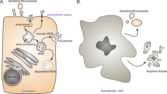
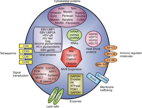
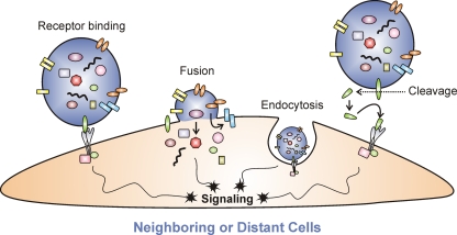
Cells secrete various membrane-enclosed microvesicles from their cell surface (shedding microvesicles) and from internal, endosome-derived membranes (exosomes). Intriguingly, these vesicles have many characteristics in common with enveloped viruses, including biophysical properties, biogenesis, and uptake by cells. Recent discoveries describing the microvesicle-mediated intercellular transfer of functional cellular proteins, RNAs, and mRNAs have revealed additional similarities between viruses and cellular microvesicles. Apparent differences include the complexity of viral entry, temporally regulated viral expression, and self-replication proceeding to infection of new cells. Interestingly, many virally infected cells secrete microvesicles that differ in content from their virion counterparts but may contain various viral proteins and RNAs. For the most part, these particles have not been analyzed for their content or functions during viral infection. However, early studies of microvesicles (L-particles) secreted from herpes simplex virus-infected cells provided the first evidence of microvesicle-mediated intercellular communication. In the case of Epstein-Barr virus, recent evidence suggests that this tumorigenic herpesvirus also utilizes exosomes as a mechanism of cell-to-cell communication through the transfer of signaling competent proteins and functional microRNAs to uninfected cells. This review focuses on aspects of the biology of microvesicles with an emphasis on their potential contributions to viral infection and pathogenesis.


Figures

Fig. 1.
Fig. 1.
Microvesicle biogenesis pathways. (A) Endocytosed proteins on the plasma membrane traffic to early endosomes where they can be sorted back to the plasma membrane or to multivesicular bodies (MVBs). MVBs contain intraluminal vesicles (ILVs) that are generated by budding from the limiting membrane of endosomes. Distinct MVB populations exist, a degradative MVB that leads to lysosomal destruction of MVB content or an exocytic pathway that traffics to the plasma membrane and, following membrane fusion, releases ILVs from the cell in the form of exosomes. Vesicles can also actively be released directly from the plasma membrane requiring a budding mechanism. These vesicles have been termed shedding microvesicles. ER, endoplasmic reticulum. (B) Dying or apoptotic cells release shedding microvesicles in the early stages of apoptosis and larger apoptotic bodies at later times that contain nuclear and cytoplasmic remnants of the degrading cell.
Fig. 2.
Fig. 2.
Molecules found in exosomes. Proteomic and biochemical analysis of purified exosomes have identified many specific proteins and RNAs present within these structures that are often in different abundances than those of their intracellular counterparts. Molecules presented here have been grouped based on functions or protein classes. vmiRNA, viral miRNA; PK, protein kinase; MHCI and MHCII, MHC class I and II, respectively; PGK1, phosphoglycerate kinase 1; MUC1, mucin 1; ARF1, ADP-ribosylation factor 1.
Fig. 3.
Fig. 3.
Potential mechanisms of microvesicle-mediated intercellular communication. Proteins present on the microvesicle surface can bind to receptors on the target cell membrane, activating signaling pathways within that cell (Receptor binding). Microvesicles may fuse with target cell membranes and release their contents at the plasma membrane (Fusion) or internal membranes following uptake into endocytic vesicles (Endocytosis). Deposited cargo can activate signaling pathways, silence target gene expression, and produce new proteins following transcription of mRNA. Microvesicles may also interact with molecules present within endocytic vesicles that induce signal transduction cascades in the absence of vesicle fusion. Proteases located in the extracellular space may cleave proteins exposed on the microvesicle surface. The potential ligands generated could bind to cell surface receptors on the target cell and initiate signaling events (Cleavage).
None
None

References

    1. Abid Hussein M. N., Boing A. N., Sturk A., Hau C. M., Nieuwland R. 2007. Inhibition of microparticle release triggers endothelial cell apoptosis and detachment. Thromb. Haemost. 98: 1096–1107 - PubMed
    1. Admyre C., Johansson S. M., Paulie S., Gabrielsson S. 2006. Direct exosome stimulation of peripheral human T cells detected by ELISPOT. Eur. J. Immunol. 36: 1772–1781 - PubMed
    1. Admyre C., et al. 2007. Exosomes with immune modulatory features are present in human breast milk. J. Immunol. 179: 1969–1978 - PubMed
    1. Ali S. A., et al. 2010. Genetic characterization of HIV type 1 Nef-induced vesicle secretion. AIDS Res. Hum. Retroviruses 26: 173–192 - PMC - PubMed
    1. Al-Nedawi K., Meehan B., Kerbel R. S., Allison A. C., Rak J. 2009. Endothelial expression of autocrine VEGF upon the uptake of tumor-derived microvesicles containing oncogenic EGFR. Proc. Natl. Acad. Sci. U. S. A. 106: 3794–3799 - PMC - PubMed

Publication types

LinkOut - more resources