. 2011 Dec;85(24):12901-9.
doi: 10.1128/JVI.06046-11. Epub 2011 Oct 5.

Rift Valley fever virus vaccine lacking the NSs and NSm genes is safe, nonteratogenic, and confers protection from viremia, pyrexia, and abortion following challenge in adult and pregnant sheep

Affiliations

Rift Valley fever virus vaccine lacking the NSs and NSm genes is safe, nonteratogenic, and confers protection from viremia, pyrexia, and abortion following challenge in adult and pregnant sheep

Brian H Bird et al. J Virol. 2011 Dec.

Abstract

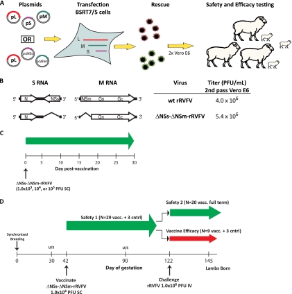
Rift Valley fever virus (RVFV) is a mosquito-borne human and veterinary pathogen causing large outbreaks of severe disease throughout Africa and the Arabian Peninsula. Safe and effective vaccines are critically needed, especially those that can be used in a targeted one-health approach to prevent both livestock and human disease. We report here on the safety, immunogenicity, and efficacy of the ΔNSs-ΔNSm recombinant RVFV (rRVFV) vaccine (which lacks the NSs and NSm virulence factors) in a total of 41 sheep, including 29 timed-pregnant ewes. This vaccine was proven safe and immunogenic for adult animals at doses ranging from 1.0 × 10(3) to 1.0 × 10(5) PFU administered subcutaneously (s.c.). Pregnant animals were vaccinated with 1.0 × 10(4) PFU s.c. at day 42 of gestation, when fetal sensitivity to RVFV vaccine-induced teratogenesis is highest. No febrile reactions, clinical illness, or pregnancy loss was observed following vaccination. Vaccination resulted in a rapid increase in anti-RVFV IgM (day 4) and IgG (day 7) titers. No seroconversion occurred in cohoused control animals. A subset of 20 ewes progressed to full-term delivery after vaccination. All lambs were born without musculoskeletal, neurological, or histological birth defects. Vaccine efficacy was assessed in 9 pregnant animals challenged at day 122 of gestation with virulent RVFV (1.0 × 10(6) PFU intravenously). Following challenge, 100% (9/9) of the animals were protected, progressed to full term, and delivered healthy lambs. As expected, all 3 sham-vaccinated controls experienced viremia, fetal death, and abortion postchallenge. These results demonstrate that the ΔNSs-ΔNSm rRVFV vaccine is safe and nonteratogenic and confers high-level protection in sheep.


Figures

Fig. 1.
Fig. 1.
(A) Schematic of the rRVFV reverse genetics rescue system. (B) Schematic of the locations of the NSs and NSm gene deletions and final endpoint titers. (C) Timeline of the pilot dose escalation study. (D) Timeline of the pregnant animal safety 1 and 2 and vaccine efficacy studies.
Fig. 2.
Fig. 2.
Pilot dose escalation serology and body temperatures. (A) Results of total anti-RVFV ELISA testing of serum, (B) Rectal temperatures. Each vaccine dosage group contained 4 animals. One animal served as a sham-inoculated control. The error bars indicate standard deviations.
Fig. 3.
Fig. 3.
Pregnant animal body temperature and viremia testing. (A) Safety 1 phase (n = 29 vaccinated animals [Vacc.] and 3 sham-vaccinated controls [Sham]). (B) Vaccine efficacy phase (n = 9 vaccinated animals and 3 sham-vaccinated controls that were challenged intravenously with 1.0 × 10⁶ PFU of virulent rRVFV at day 80 postvaccination [day 122 gestation]). Temperatures and serum specimens for qRT-PCR testing were obtained during the early morning from each animal. The error bars indicate standard deviations of ± 1.0.
Fig. 4.
Fig. 4.
Pregnant animal anti-RVFV serology results. (A) Safety 1 phase total anti-RVFV ELISA testing (n = 29 vaccinated and 3 sham vaccinated) of sheep. After day 82 p.v., the data represent the 20 vaccinated animals that comprised the safety 2 arm of the trial. (B) Total anti-RVFV ELISA results for animals (n = 9 vaccinated and 3 sham vaccinated) that were challenged intravenously with 1.0 × 10⁶ PFU of virulent rRVFV at day 82 postvaccination. The days postchallenge are indicated by gray shading. Note the rapid rise in anti-RVFV IgG among vaccinated animals compared with the sham-vaccinated controls. (C) Results of PRNT80 testing during the safety 1 phase (n = 29 vaccinated animals and 3 sham-vaccinated controls). (D) Results of 3-way (NP-NSs-NSm) DIVA ELISA testing at day 54 postvaccination or day 37 postchallenge. The data represent the results from the vaccine efficacy phase of the experiment (n = 9 vaccinated animals and 3 sham-vaccinated control animals). Serum was assayed in 2-fold dilutions from 1:100 to 1:800. The error bars indicate standard deviations of ± 1.0.
Fig. 5.
Fig. 5.
Gross and histological examination of lamb tissues. (A) Coronal brain sections at the level of the lateral ventricles and hypothalamus obtained from lambs born to the safety 2 phase animals (n = 20 vaccinated ewes and 22 lambs). Note the presence of normal ventricle size and architecture. Also note that slight normal variation can be observed in the color and ventricle shape between the brain sections due to differences in the exact plane of transection. Two brain sections obtained from similarly aged (1- to 5-day-old) normal lambs are shown for comparison (lower right, Cntrl1 and Cntrl2). (B) Anti-RVFV immunohistochemistry. (Top row) Specimens (×20 magnification) from an aborted RVFV-positive (number 104) fetus depicting liver, cerebrum, and cerebellum (left to right, respectively). Marked diffuse necrosis and antigen staining (red to brown pigment) are apparent in the liver tissue (×40 inset); focal antigen and serum staining can be observed in the cerebrum and cerebellum tissues. (Bottom row) Specimens from a lamb (number 11) born postchallenge to a previously vaccinated ewe; histologically normal and RVFV antigen-negative liver, cerebrum, and cerebellum tissues are shown (left to right, respectively).

References

    1. Anyangu A. S., et al. 2010. Risk factors for severe Rift Valley fever infection in Kenya, 2007. Am. J. Trop. Med. Hyg. 83:14–21 - PMC - PubMed
    1. Beaty B. J., Rozhon E. J., Gensemer P., Bishop D. H. 1981. Formation of reassortant bunyaviruses in dually infected mosquitoes. Virology 111:662–665 - PubMed
    1. Billecocq A., et al. 2004. NSs protein of Rift Valley fever virus blocks interferon production by inhibiting host gene transcription. J. Virol. 78:9798–9806 - PMC - PubMed
    1. Bird B. H., et al. 2008. Rift valley fever virus lacking the NSs and NSm genes is highly attenuated, confers protective immunity from virulent virus challenge, and allows for differential identification of infected and vaccinated animals. J. Virol. 82:2681–2691 - PMC - PubMed
    1. Bird B. H., Albarino C. G., Nichol S. T. 2007. Rift Valley fever virus lacking NSm proteins retains high virulence in vivo and may provide a model of human delayed onset neurologic disease. Virology 362:10–15 - PubMed

MeSH terms

LinkOut - more resources