. 2011 Dec;85(24):13310-21.
doi: 10.1128/JVI.05728-11. Epub 2011 Oct 5.

Infection with seasonal influenza virus elicits CD4 T cells specific for genetically conserved epitopes that can be rapidly mobilized for protective immunity to pandemic H1N1 influenza virus

Affiliations

Infection with seasonal influenza virus elicits CD4 T cells specific for genetically conserved epitopes that can be rapidly mobilized for protective immunity to pandemic H1N1 influenza virus

Shabnam Alam et al. J Virol. 2011 Dec.

Abstract

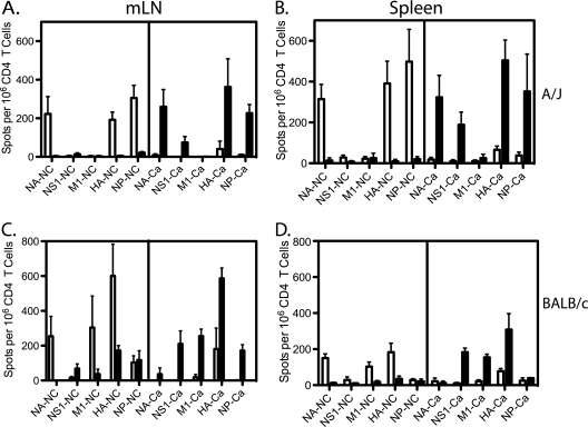
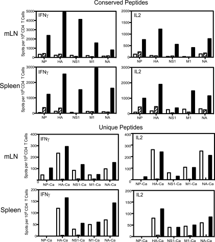
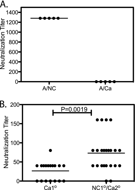
In recent years, influenza viruses with pandemic potential have been a major concern worldwide. One unresolved issue is how infection or vaccination with seasonal influenza virus strains influences the ability to mount a protective immune response to novel pandemic strains. In this study, we developed a mouse model of primary and secondary influenza infection by using a widely circulating seasonal H1N1 virus and the pandemic strain of H1N1 that emerged in Mexico in 2009, and we evaluated several key issues. First, using overlapping peptide libraries encompassing the entire translated sequences of 5 major influenza virus proteins, we assessed the specificity of CD4 T cell reactivity toward epitopes conserved among H1N1 viruses or unique to the seasonal or pandemic strain by enzyme-linked immunospot (ELISpot) assays. Our data show that CD4 T cells reactive to both virus-specific and genetically conserved epitopes are elicited, allowing separate tracking of these responses. Populations of cross-reactive CD4 T cells generated from seasonal influenza infection were found to expand earlier after secondary infection with the pandemic H1N1 virus than CD4 T cell populations specific for new epitopes. Coincident with this rapid CD4 T cell response was a potentiated neutralizing-antibody response to the pandemic strain and protection from the pathological effects of infection with the pandemic virus. This protection was not dependent on CD8 T cells. Together, our results indicate that exposure to seasonal vaccines and infection elicits CD4 T cells that promote the ability of the mammalian host to mount a protective immune response to pandemic strains of influenza virus.


Figures

Fig. 1.
Fig. 1.
A/J mice are much more susceptible to the A/California/04/09 (A/Ca) virus than BALB/c mice. (A) Weight loss was recorded for 12 days after mice were infected with different doses (circles, 15,000 EID50; squares, 125,000 EID50) of the A/California/04/09 virus via intranasal inhalation. Open symbols, A/J mice; filled symbols, BALB/c mice. Means for 5 mice in each group ± standard errors of the means are shown. (B) Mortality of A/J mice (filled circles) versus mortality of BALB/c mice. Data for mice of the same strain receiving different doses of virus were combined. Results for a total of 10 mice of each strain are shown. Surviving A/J mice were euthanized at day 12, because they had lost 30% of their body weight. Dotted lines represent 10% weight loss (A) or 50% survival (B).
Fig. 2.
Fig. 2.
Previous exposure to the seasonal A/New Caledonia/20/99 virus protects against the A/California/04/09 (A/Ca) virus. Naïve mice (open symbols) or mice previously primed by infection with 50,000 EID50 of the A/New Caledonia/20/99 virus (filled symbols) were infected intranasally with 15,000 EID50 of the A/California/04/09 virus. (A) Body weight loss was recorded for 12 days. Values are mean percentages of starting body weight for the 5 mice in each group ± standard errors of the means. (B) Virus titers in the lung at day 5 after infection with the A/California/04/09 virus were measured using a plaque assay. Means ± standard errors of the means are shown. (C) Survival was recorded after infection with the A/California/04/09 virus. Remaining mice were euthanized at day 12, because they had lost 30% of their body weight, and were included in mortality data. Dotted lines represent 10% weight loss (A) or 50% survival (C).
Fig. 3.
Fig. 3.
CD8 T cells are not needed for protection after secondary infection. A/J mice were primed with 50,000 EID50 of the A/New Caledonia/20/99 virus and were rested for 8 to 9 weeks. For CD8 depletion in vivo, 200 μg of an anti-CD8 or isotype control IgG2b antibody was injected intraperitoneally 2 days prior to challenge, on the day of challenge, and 2 days after challenge with 15,000 EID50 of the A/California/04/09 (A/Ca) virus. Mice were observed daily for weight loss and survival. Means ± standard errors of the means are shown for 3 mice in each group. Shown are the primary response to the A/California/04/09 virus (open circles) and the secondary responses to the A/California/04/09 virus in anti-CD8-treated mice (filled squares) and isotype control IgG2b-treated mice (filled triangles). Dotted lines mark 10% weight loss.
Fig. 4.
Fig. 4.
Rapid CD4 T cell response at day 5 assessed by using total peptide pools in A/J mice. (A) Experimental design for effect of immunological memory induced by seasonal H1N1 virus infection. A/J mice were infected intranasally with 50,000 EID50 of A/New Caledonia/20/99 (A/NC) and were then rested for 8 to 9 weeks. One cohort of these mice was not further challenged, and a second cohort was challenged with 15,000 EID50 of A/California/04/09 (A/Ca) (secondary challenge). A cohort of naïve mice was infected with the A/California/04/09 virus in order to analyze the responses from a primary infection in parallel. (B) At day 5 after infection with the A/California/04/09 virus, CD4 T cells were isolated from the mediastinal lymph nodes (mLN) or the spleen. Open bars, primary response to A/California/04/09; hatched bars, residual CD4 T cells from memory without challenge; filled bars, secondary response. To visualize all influenza virus-reactive cells, “complete” peptide pools comprising overlapping peptides representing sequences from A/New Caledonia/20/99 were used to stimulate CD4 T cells. The proteins represented, indicated below the bar graphs, were incubated with syngeneic APC and CD4 T cells overnight. Cells producing IFN-γ (left) or IL-2 (right) were quantified by ELISpot assays. The data are presented as spots per million CD4 T cells.
Fig. 5.
Fig. 5.
Reactivity of CD4 T cells responding to genetically conserved peptides upon infection with a seasonal or pandemic influenza virus. A/J (A and B) or BALB/c (C and D) mice were infected intranasally with either 50,000 EID50 of the A/New Caledonia/20/99 strain (open bars) or 15,000 EID50 of the A/California/04/09 strain (filled bars). At days 8 to 10 postinfection, CD4 T cells were isolated from mediastinal lymph nodes (mLN) or spleens and were incubated with syngeneic APC and pools of synthetic peptides conserved in A/New Caledonia/20/99 and A/California/04/09 (listed in Table S1 in the supplemental material), derived from the seasonal influenza virus proteins given beneath each panel. Peptide-specific IFN-γ-producing cells were quantified by ELISpot assays, and the data are presented as responders per million CD4 T cells.
Fig. 6.
Fig. 6.
CD4 T cells from seasonal and pandemic influenza viruses can be distinguished. A/J (A and B) or BALB/c (C and D) mice were infected intranasally with either 50,000 EID50 of the A/New Caledonia/20/99 strain (open bars) or 15,000 EID50 of the A/California/04/09 strain (filled bars). At days 8 to 10 postinfection, CD4 T cells were isolated from mediastinal lymph nodes or spleens and were incubated with syngeneic APC and the indicated peptides (listed in Table S2 in the supplemental material). Pools of peptides representing unique sequences from A/New Caledonia/20/99 (NC) or A/California/04/09 (Ca) are indicated beneath each panel. Peptide-specific and IFN-γ-producing cells were quantified by ELISpot assays, and the data are presented as responders per million CD4 T cells.
Fig. 7.
Fig. 7.
Analysis of the specificity of the early CD4 T cell response in secondary challenge. Using the protocol outlined in Fig. 4A, at day 5 after A/California/04/09 infection, CD4 T cells were isolated from the mediastinal lymph nodes (mLN) or spleen and were assayed for reactivity to the indicated pools of peptides representing either conserved or virus-unique sequences. Open bars, primary responses to A/California/04/09; hatched bars, memory CD4 T cells; filled bars, secondary responses. Either “conserved” or “unique” peptide pools, representing the particular viral proteins given beneath each bar graph, were used to stimulate CD4 T cells. Cells producing IFN-γ (left) or IL-2 (right) were quantified by ELISpot assays, and the data are presented as spots per million CD4 T cells.
Fig. 8.
Fig. 8.
Infection with seasonal influenza virus potentiates the B cell response to the pandemic strain. (A) Lack of antibody cross-reactivity between A/New Caledonia/20/99 (A/NC) and A/California/04/09 (A/Ca). Serum samples collected from A/J mice 8 weeks after infection with A/New Caledonia/20/99 were assayed for reactivity by a microneutralization assay using either A/New Caledonia/20/99 or A/California/04/09, as indicated. (B) Accelerated neutralizing antibody response to the A/California/04/09 virus by earlier priming with A/New Caledonia/20/99 (NC1o/Ca2o). Primed mice were rested 8 to 9 weeks and were then challenged by infection with 15,000 EID50 of A/California/04/09. Control mice (Ca1o) were not previously primed but were infected with 15,000 EID50 of A/California/04/09. Sera were collected at day 5 after A/California/04/09 infection and were tested by a microneutralization assay with A/California/04/09. Statistical significance was evaluated using an unpaired Student t test.

References

    1. Alberts R., et al. 2010. Gene expression changes in the host response between resistant and susceptible inbred mouse strains after influenza A infection. Microbes Infect. 12:309–318 - PubMed
    1. Albright F. S., Orlando P., Pavia A. T., Jackson G. G., Cannon Albright L. A. 2008. Evidence for a heritable predisposition to death due to influenza. J. Infect. Dis. 197:18–24 - PubMed
    1. Ansaldi F., Durando P., Icardi G. 2011. Intradermal influenza vaccine and new devices: a promising chance for vaccine improvement. Expert Opin. Biol. Ther. 11:415–427 - PubMed
    1. Bautista E., et al. 2010. Clinical aspects of pandemic 2009 influenza A (H1N1) virus infection. N. Engl. J. Med. 362:1708–1719 - PubMed
    1. Berard M., Tough D. F. 2002. Qualitative differences between naive and memory T cells. Immunology 106:127–138 - PMC - PubMed

Publication types