. 2011 Nov 25;286(47):40509-19.
doi: 10.1074/jbc.M111.287201. Epub 2011 Oct 5.

PEX5 protein binds monomeric catalase blocking its tetramerization and releases it upon binding the N-terminal domain of PEX14

Affiliations

PEX5 protein binds monomeric catalase blocking its tetramerization and releases it upon binding the N-terminal domain of PEX14

Marta O Freitas et al. J Biol Chem. .

Abstract

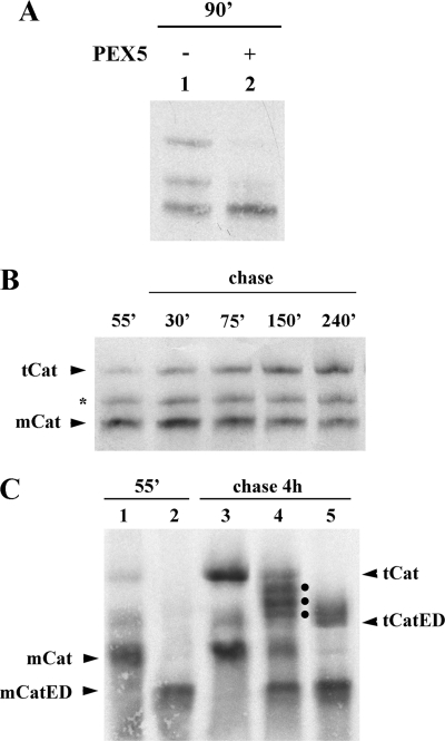
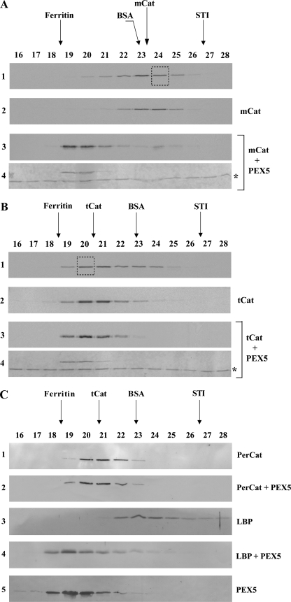
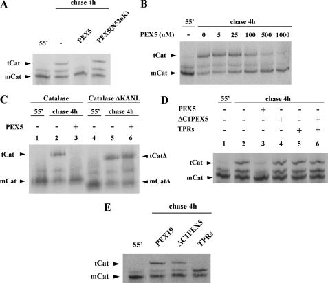
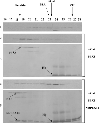
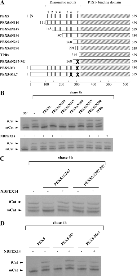
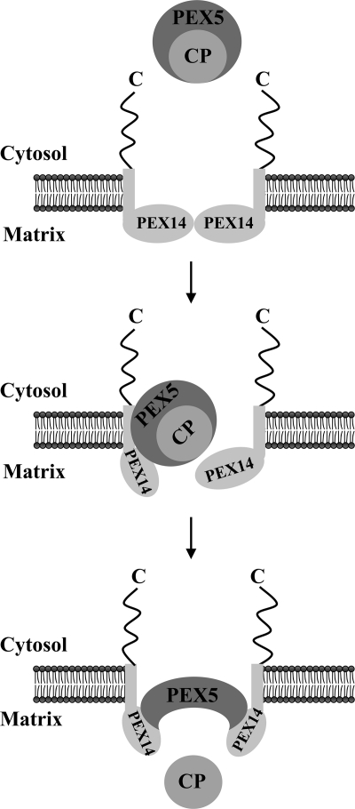
Newly synthesized peroxisomal matrix proteins are targeted to the organelle by PEX5. PEX5 has a dual role in this process. First, it acts as a soluble receptor recognizing these proteins in the cytosol. Subsequently, at the peroxisomal docking/translocation machinery, PEX5 promotes their translocation across the organelle membrane. Despite significant advances made in recent years, several aspects of this pathway remain unclear. Two important ones regard the formation and disruption of the PEX5-cargo protein interaction in the cytosol and at the docking/translocation machinery, respectively. Here, we provide data on the interaction of PEX5 with catalase, a homotetrameric enzyme in its native state. We found that PEX5 interacts with monomeric catalase yielding a stable protein complex; no such complex was detected with tetrameric catalase. Binding of PEX5 to monomeric catalase potently inhibits its tetramerization, a property that depends on domains present in both the N- and C-terminal halves of PEX5. Interestingly, the PEX5-catalase interaction is disrupted by the N-terminal domain of PEX14, a component of the docking/translocation machinery. One or two of the seven PEX14-binding diaromatic motifs present in the N-terminal half of PEX5 are probably involved in this phenomenon. These results suggest the following: 1) catalase domain(s) involved in the interaction with PEX5 are no longer accessible upon tetramerization of the enzyme; 2) the catalase-binding interface in PEX5 is not restricted to its C-terminal peroxisomal targeting sequence type 1-binding domain and also involves PEX5 N-terminal domain(s); and 3) PEX14 participates in the cargo protein release step.


Figures

FIGURE 1.
FIGURE 1.
35S-Labeled catalase tetramerizes in vitro. A, human catalase was synthesized in vitro in a rabbit reticulocyte lysate for 90 min at 30 °C in the absence or presence of 1 μm human PEX5, as indicated, and analyzed by native-PAGE/autoradiography. B, 35S-labeled catalase was synthesized for 55 min. After adding cycloheximide, an aliquot was removed and frozen in liquid N2 (lane 55′). The remainder of the reaction was then incubated at 30 °C, and aliquots were removed and frozen at the indicated time points. The samples were subjected to native-PAGE/autoradiography. The protein bands labeled with mCat and tCat correspond to the monomeric and tetrameric forms of catalase; the band labeled with an asterisk probably represents dimeric catalase (see text for details). C, catalase and a mutant version of it possessing two acidic amino acid residues at the C terminus (CatED) were synthesized in vitro for 55 min and supplemented with cycloheximide (lanes 1 and 2, respectively). Aliquots of each reaction were then combined and incubated for 4 h at 30 °C (lane 4) or incubated individually under the same conditions (lanes 3 and 5 for catalase and CatED, respectively), and subjected to native PAGE/autoradiography. Note that this gel was run for 2.5 h to improve separation of tetramers. Longer electrophoretic runs also result in more diffuse bands. The dots in lane 4 indicate the three expected heterotetramers. mCatED and tCatED indicate the monomeric and tetrameric forms of CatED, respectively.
FIGURE 2.
FIGURE 2.
PEX5 binds monomeric catalase. A, 35S-labeled catalase was synthesized in vitro for 55 min and subjected to SEC. Radiolabeled mCat eluting in fraction 24 of this chromatography (panel 1, boxed lane) was then subjected to a second SEC either alone (panel 2) or after receiving 1 μm recombinant PEX5 (panels 3 and 4). Fractions were collected and subjected to SDS-PAGE/Western blotting. Autoradiographs (panels 1–3) and the Ponceau S-stained membrane showing PEX5 (panel 4) are presented. No recombinant PEX5 or 35S-labeled catalase were detected in the void volume of this column (fractions 14 and 15; not shown). The asterisk marks bovine serum albumin added to chromatography fractions before precipitation to control protein recoveries. B, 35S-labeled catalase, synthesized in vitro for 55 min and incubated for 4 h at 30 °C in the presence of cycloheximide, was subjected to SEC. Radiolabeled tCat eluting in fraction 20 (panel 1, boxed lane) was then subjected to a second SEC either alone (panel 2) or after receiving 1 μm recombinant PEX5 (panels 3 and 4). Fractions were processed as described above. Autoradiographs (panels 1–3) and the Ponceau S-stained membrane (panel 4) are presented. C, soluble proteins from mouse liver peroxisomes were incubated either with recombinant PEX5 or buffer alone and subjected to SEC. Fractions were subjected to SDS-PAGE/Western blotting using antibodies directed to catalase (PerCat) or L-bifunctional protein. Immunoblots (panels 1–4) and a Ponceau S-stained membrane showing PEX5 (panel 5) are presented. Note that PEX5, a monomeric 70-kDa protein in solution, displays an abnormal behavior upon SEC because a major fraction of its polypeptide chain is natively unfolded (52).
FIGURE 3.
FIGURE 3.
PEX5 inhibits catalase tetramerization. A, 35S-labeled catalase was synthesized in vitro for 55 min (lane 55′) and chased for 4 h in the absence (lane −) or presence of 1 μm of the indicated recombinant proteins. B, same as in A, but using the indicated concentrations of PEX5. C, catalase (lanes 1–3) and a truncated version of it lacking the PTS1 signal (catalaseΔKANL; lanes 4–6) were synthesized for 55 min and chased in the absence (lanes 2 and 5) or presence of 1 μm PEX5 (lanes 3 and 6). D, 35S-labeled catalase was synthesized in vitro for 55 min (lane 1) and chased in the absence (lane 2) or presence of 1 μm of the indicated recombinant proteins (lanes 3–6). ΔC1PEX5 and TPRs, recombinant proteins comprising the N- and C-terminal half of PEX5, respectively. E, same as in A, but using 200 μm of the indicated recombinant proteins. Samples were analyzed by native-PAGE/autoradiography. Note that the gel shown in C was run for 2.5 h. mCat and tCat, monomeric and tetrameric versions of catalase, respectively; mCatΔ and tCatΔ, monomeric and tetrameric forms of catalaseΔKANL, respectively.
FIGURE 4.
FIGURE 4.
N-terminal domain of PEX14 disrupts the mCat-PEX5 interaction. 35S-Labeled mCat was purified by SEC (panel 1, fraction 24), supplemented with 1 μm recombinant PEX5 and incubated for 30 min at room temperature to generate the PEX5-mCat protein complex. Half of this sample was analyzed directly by SEC (panels 2 and 3). The other half received recombinant NDPEX14 (15 μm) 30 min before chromatography (panels 4 and 5). Fractions were subjected to SDS-PAGE and blotted onto a nitrocellulose membrane. Autoradiographs (panels 1, 2, and 4) and the Ponceau S-stained membranes (panels 3 and 5) are presented. Hb, hemoglobin from the reticulocyte lysate that co-purified with mCat in the first SEC.
FIGURE 5.
FIGURE 5.
PEX5 diaromatic motifs involved in the NDPEX14-induced disruption of the mCat-PEX5 interaction. A, schematic representation of recombinant PEX5 proteins. The diaromatic motifs in the N-terminal half of PEX5 are numbered 1–7. Replacement of tryptophan and phenylalanine/tyrosine residues by alanines in these motifs is indicated by X. B–D, 35S-labeled catalase was synthesized in vitro for 55 min (lane 55′) and chased for 4 h in the absence (lane −) or presence of 1 μm of the indicated recombinant PEX5 proteins alone or together with NDPEX14 (30 μm). Samples were analyzed by native-PAGE/autoradiography.
FIGURE 6.
FIGURE 6.
Role of PEX14 in the release of cargo proteins into the peroxisomal matrix. A newly synthesized cargo protein (CP) is recognized by PEX5 in the cytosol. This protein complex then docks at and becomes inserted into the peroxisomal DTM of which only PEX14 is shown for simplicity. The DTM component(s) providing the docking site for the PEX5-cargo protein complex have not been unambiguously identified yet. As discussed elsewhere, two strong candidates for this role are PEX13 (86) and PEX14 itself (87, 88). Note that PEX13 may also participate in the cargo-release step (66). The multiple interactions of PEX5 with the N-terminal domain of the several PEX14 molecules present in the DTM ultimately trigger the release of the cargo into the peroxisomal matrix.

Similar articles

Cited by

References

    1. Grou C. P., Carvalho A. F., Pinto M. P., Alencastre I. S., Rodrigues T. A., Freitas M. O., Francisco T., Sá-Miranda C., Azevedo J. E. (2009) Cell. Mol. Life Sci. 66, 254–262 - PMC - PubMed
    1. Lanyon-Hogg T., Warriner S. L., Baker A. (2010) Biol. Cell 102, 245–263 - PubMed
    1. Ma C., Subramani S. (2009) IUBMB Life 61, 713–722 - PMC - PubMed
    1. Wolf J., Schliebs W., Erdmann R. (2010) FEBS J. 277, 3268–3278 - PubMed
    1. Brocard C., Hartig A. (2006) Biochim. Biophys. Acta 1763, 1565–1573 - PubMed

Publication types

MeSH terms