Case Reports
. 2011 Dec;96(12):E2078-87.
doi: 10.1210/jc.2011-1401. Epub 2011 Oct 5.

Hereditary pituitary hyperplasia with infantile gigantism

Affiliations
Case Reports

Hereditary pituitary hyperplasia with infantile gigantism

Sven Gläsker et al. J Clin Endocrinol Metab. 2011 Dec.

Abstract

Context: We report hereditary pituitary hyperplasia.

Objective: The objective of the study was to describe the results of the clinical and laboratory analysis of this rare instance of hereditary pituitary hyperplasia.

Design: The study is a retrospective analysis of three cases from one family.

Setting: The study was conducted at the National Institutes of Health, a tertiary referral center.

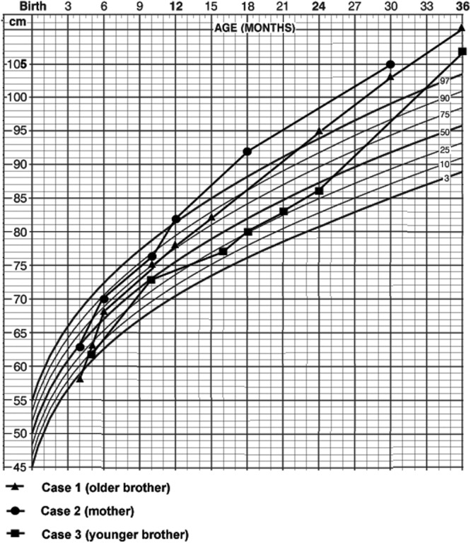
Patients: A mother and both her sons had very early-onset gigantism associated with high levels of serum GH and prolactin.

Interventions: The condition was treated by total hypophysectomy.

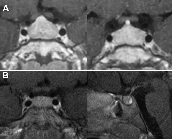
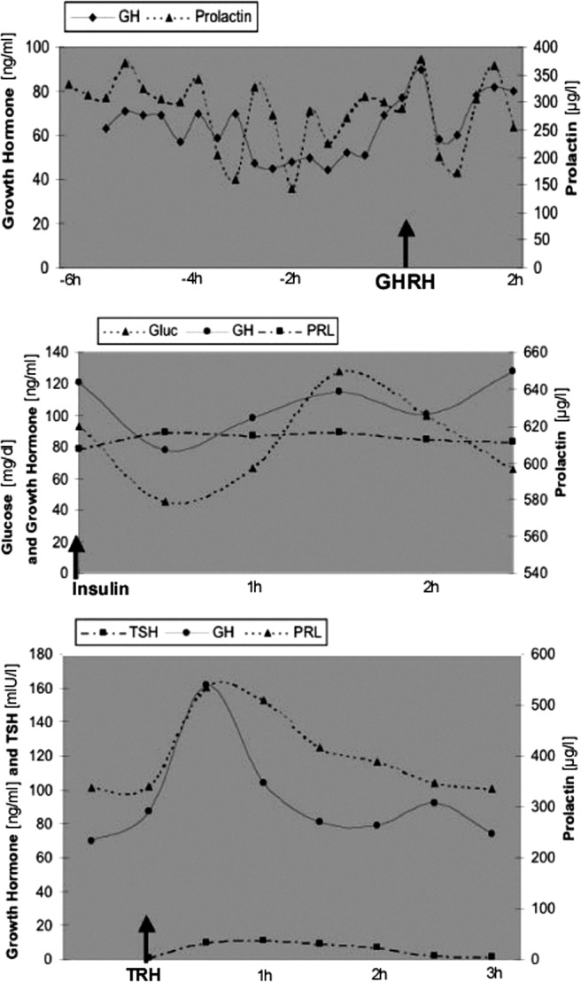
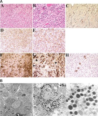
Main outcome measure(s): We performed clinical, pathological, and molecular evaluations, including evaluation basal and provocative endocrine testing, neuroradiological assessment, and assessment of the pituitary tissue by microscopic evaluation, immunohistochemistry, and electron microscopy.

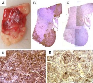
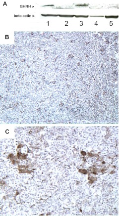
Results: All three family members had very early onset of gigantism associated with abnormally high serum levels of GH and prolactin. Serum GHRH levels were not elevated in either of the boys. The clinical, radiographic, surgical, and histological findings indicated mammosomatotroph hyperplasia. The pituitary gland of both boys revealed diffuse mammosomatotroph hyperplasia of the entire pituitary gland without evidence of adenoma. Prolactin and GH were secreted by the same cells within the same secretory granules. Western blot and immunohistochemistry demonstrated expression of GHRH in clusters of cells distributed throughout the hyperplastic pituitary of both boys.

Conclusions: This hereditary condition seems to be a result of embryonic pituitary maldevelopment with retention and expansion of the mammosomatotrophs. The findings suggest that it is caused by paracrine or autocrine pituitary GHRH secretion during pituitary development.


Figures

Fig. 1.
Fig. 1.
Growth curves of the mother and her sons.
Fig. 2.
Fig. 2.
Preoperative contrast-enhanced MR imaging scans of the older brother (case 1) demonstrating symmetric enlargement of the pituitary (A) and the younger brother (case 3) demonstrating a slightly enlarged, symmetric gland (B). The posterior lobe is prominently seen. No focus suggestive of an adenoma is present in either scan.
Fig. 3.
Fig. 3.
Results of basal and provocative biochemical testing in case 1. Top panel, GH and PRL levels increase immediately after GHRH stimulation. Middle panel, GH reaches its nadir of 80 ng/ml 30 min after the insulin challenge, when the glucose level reaches 40 mg/dl. Bottom panel, Serum GH and PRL peak approximately 30 min after TRH injection.
Fig. 4.
Fig. 4.
A, Pathological features of case 1. Panels A and B, Hematoxylin and eosin sections of the pituitary lesion at medium (×200) and high-power (×400) magnifications, respectively. Enlarged acini are nearly entirely filled with acidophilic endocrine cells. Panel C, Reticulin stain (×200) demonstrates acinar expansion caused by pituitary hyperplasia. Panels D and E, Immunohistochemical staining with GH (×200 and ×400). Panels F and G, Immunohistochemical staining with PRL (×200 and 400, respectively), demonstrates perinuclear inclusion bodies (arrows in G). Panel H, Immunohistochemical staining with ACTH through the same sections as panels D–G shows rare positive cells scattered among the PRL- and ACTH-positive cells. B, Ultrastructural features of case 1. Immunolabeling of pituitary hyperplasia with antibodies directed against GH (small gold beads, ∼10 nm in diameter) and PRL (large gold beads, ∼20 nm in diameter). Most cells are positive for PRL and GH. Within the individual cells, numerous neuroendocrine granules contain both PRL and GH, and occasional granules contain PRL or GH only. In A, black bar indicates 5 μm distance; B, 1 μm distance; and C, 0.5 μm.
Fig. 5.
Fig. 5.
A, The surgical specimen of case 3 consisted of homogeneous tissue with no evidence of adenoma. Immunohistochemistry for GH in ×1 (B) and ×40 (D) magnification revealed staining of the entire specimen with accentuation in the areas that look white on macroscopic examination (A). Immunohistochemistry for PRL (C and E) also revealed staining of the entire specimen; however, the areas that appeared pink on macroscopic examination were accentuated (C: ×1, E: ×40 magnification).
Fig. 6.
Fig. 6.
A, Western blot revealed that GHRH was expressed in two separate samples of anterior pituitary gland (lanes 1 and 3) from case 3. A small piece of posterior lobe, which had been attached to the surgical specimen, did not show expression of GHRH (lane 2). As negative controls we used a pituitary specimen from a patient with sporadic mammosomatotroph hyperplasia (McCune-Albright, lane 4) and a piece of normal brain (lane 5). B and C, Immunohistochemistry with anti-GHRH confirms the expression of GHRH by a subset of cells distributed within the tissue surrounded by the majority of the cells that do not express GHRH. The positive cells were assembled in groups (B, ×10; C, ×40 magnification).

Similar articles

Cited by

  • Pituitary gigantism: update on molecular biology and management.
    Lodish MB, Trivellin G, Stratakis CA. Lodish MB, et al. Curr Opin Endocrinol Diabetes Obes. 2016 Feb;23(1):72-80. doi: 10.1097/MED.0000000000000212. Curr Opin Endocrinol Diabetes Obes. 2016. PMID: 26574647 Free PMC article. Review.
  • Genetics of Acromegaly and Gigantism.
    Bogusławska A, Korbonits M. Bogusławska A, et al. J Clin Med. 2021 Mar 29;10(7):1377. doi: 10.3390/jcm10071377. J Clin Med. 2021. PMID: 33805450 Free PMC article. Review.
  • Is IGSF1 involved in human pituitary tumor formation?
    Faucz FR, Horvath AD, Azevedo MF, Levy I, Bak B, Wang Y, Xekouki P, Szarek E, Gourgari E, Manning AD, de Alexandre RB, Saloustros E, Trivellin G, Lodish M, Hofman P, Anderson YC, Holdaway I, Oldfield E, Chittiboina P, Nesterova M, Biermasz NR, Wit JM, Bernard DJ, Stratakis CA. Faucz FR, et al. Endocr Relat Cancer. 2015 Feb;22(1):47-54. doi: 10.1530/ERC-14-0465. Epub 2014 Dec 19. Endocr Relat Cancer. 2015. PMID: 25527509 Free PMC article.
  • Somatic GNAS mutation causes widespread and diffuse pituitary disease in acromegalic patients with McCune-Albright syndrome.
    Vortmeyer AO, Gläsker S, Mehta GU, Abu-Asab MS, Smith JH, Zhuang Z, Collins MT, Oldfield EH. Vortmeyer AO, et al. J Clin Endocrinol Metab. 2012 Jul;97(7):2404-13. doi: 10.1210/jc.2012-1274. Epub 2012 May 7. J Clin Endocrinol Metab. 2012. PMID: 22564667 Free PMC article.
  • Gigantism and acromegaly due to Xq26 microduplications and GPR101 mutation.
    Trivellin G, Daly AF, Faucz FR, Yuan B, Rostomyan L, Larco DO, Schernthaner-Reiter MH, Szarek E, Leal LF, Caberg JH, Castermans E, Villa C, Dimopoulos A, Chittiboina P, Xekouki P, Shah N, Metzger D, Lysy PA, Ferrante E, Strebkova N, Mazerkina N, Zatelli MC, Lodish M, Horvath A, de Alexandre RB, Manning AD, Levy I, Keil MF, Sierra Mde L, Palmeira L, Coppieters W, Georges M, Naves LA, Jamar M, Bours V, Wu TJ, Choong CS, Bertherat J, Chanson P, Kamenický P, Farrell WE, Barlier A, Quezado M, Bjelobaba I, Stojilkovic SS, Wess J, Costanzi S, Liu P, Lupski JR, Beckers A, Stratakis CA. Trivellin G, et al. N Engl J Med. 2014 Dec 18;371(25):2363-74. doi: 10.1056/NEJMoa1408028. Epub 2014 Dec 3. N Engl J Med. 2014. PMID: 25470569 Free PMC article.

References

    1. Lloyd RV, Jin L, Chang A, Kulig E, Camper SA, Ross BD, Downs TR, Frohman LA. 1992. Morphologic effects of hGRH gene expression on the pituitary, liver, and pancreas of MT-hGRH transgenic mice. An in situ hybridization analysis. Am J Pathol 141:895–906 - PMC - PubMed
    1. Mayo KE, Hammer RE, Swanson LW, Brinster RL, Rosenfeld MG, Evans RM. 1988. Dramatic pituitary hyperplasia in transgenic mice expressing a human growth hormone-releasing factor gene. Mol Endocrinol 2:606–612 - PubMed
    1. Asa SL, Kovacs K, Stefaneanu L, Horvath E, Billestrup N, Gonzalez-Manchon C, Vale W. 1990. Pituitary mammosomatotroph adenomas develop in old mice transgenic for growth hormone-releasing hormone. Proc Soc Exp Biol Med 193:232–235 - PubMed
    1. Espiner EA, Carter TA, Abbott GD, Wrightson P. 1981. Pituitary gigantism in a 31 month old girl: endocrine studies and successful response to hypophysectomy. J Endocrinol Invest 4:445–450 - PubMed
    1. Penny ES, Penman E, Price J, Rees LH, Sopwith AM, Wass JA, Lytras N, Besser GM. 1984. Circulating growth hormone releasing factor concentrations in normal subjects and patients with acromegaly. Br Med J (Clin Res Ed) 289:453–455 - PMC - PubMed

Publication types