. 2012 Feb;40(3):1318-30.
doi: 10.1093/nar/gkr812. Epub 2011 Oct 5.

Three critical hydrogen bonds determine the catalytic activity of the Diels-Alderase ribozyme

Affiliations

Three critical hydrogen bonds determine the catalytic activity of the Diels-Alderase ribozyme

Stefanie Kraut et al. Nucleic Acids Res. 2012 Feb.

Abstract

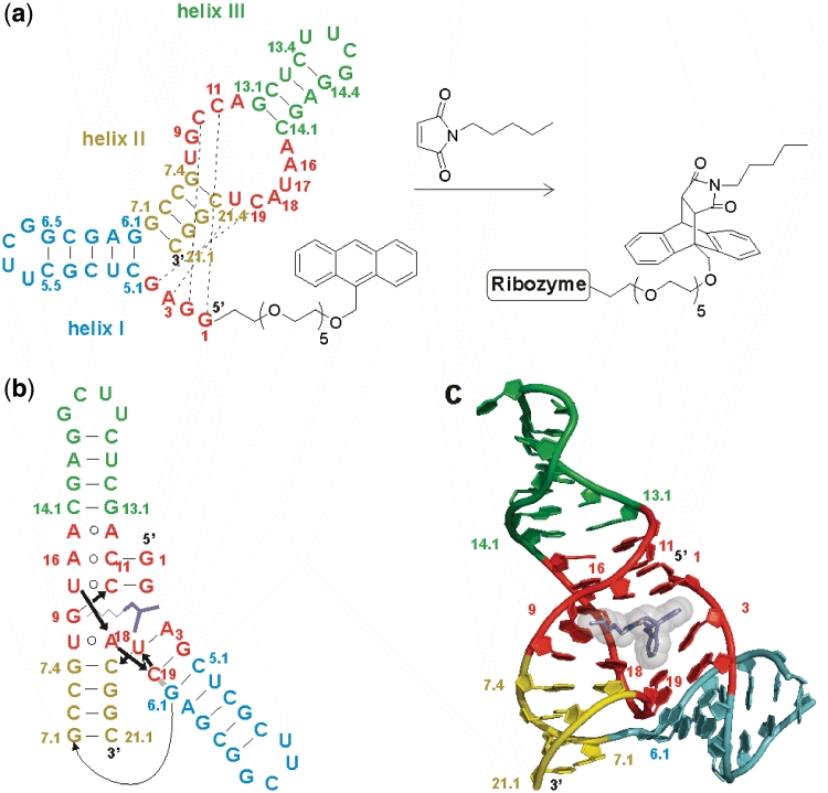
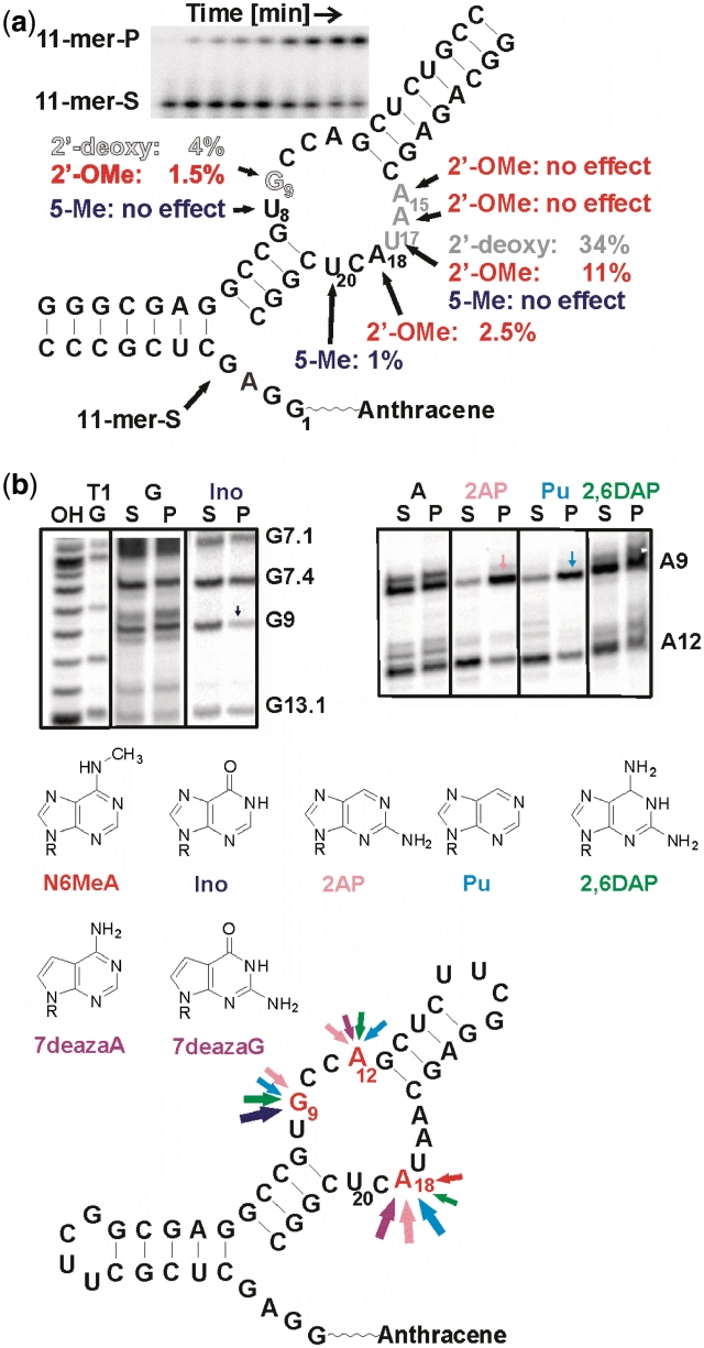
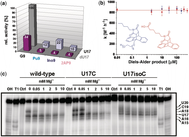
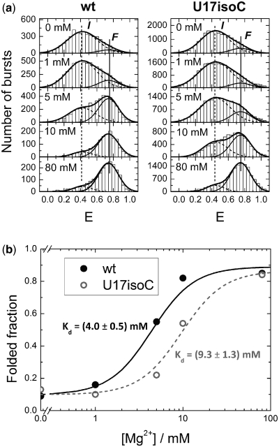
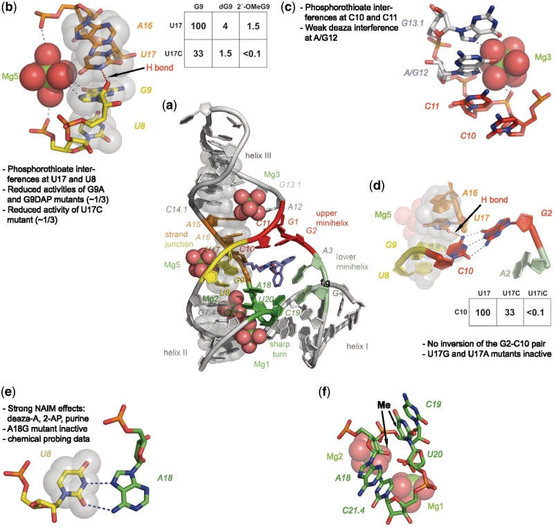
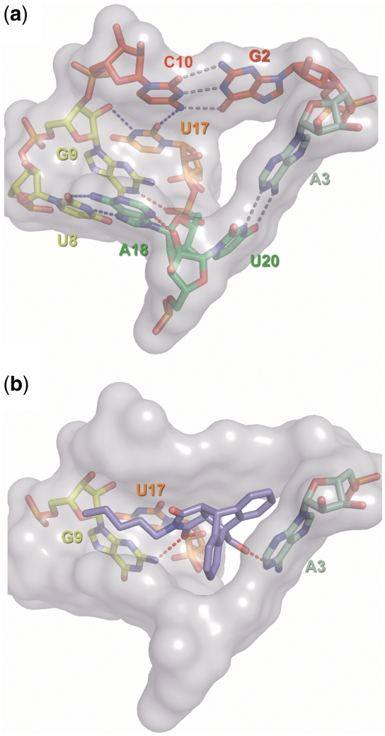
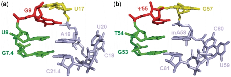
Compared to protein enzymes, our knowledge about how RNA accelerates chemical reactions is rather limited. The crystal structures of a ribozyme that catalyzes Diels-Alder reactions suggest a rich tertiary architecture responsible for catalysis. In this study, we systematically probe the relevance of crystallographically observed ground-state interactions for catalytic function using atomic mutagenesis in combination with various analytical techniques. The largest energetic contribution apparently arises from the precise shape complementarity between transition state and catalytic pocket: A single point mutant that folds correctly into the tertiary structure but lacks one H-bond that normally stabilizes the pocket is completely inactive. In the rate-limiting chemical step, the dienophile is furthermore activated by two weak H-bonds that contribute ∼7-8 kJ/mol to transition state stabilization, as indicated by the 25-fold slower reaction rates of deletion mutants. These H-bonds are also responsible for the tight binding of the Diels-Alder product by the ribozyme that causes product inhibition. For high catalytic activity, the ribozyme requires a fine-tuned balance between rigidity and flexibility that is determined by the combined action of one inter-strand H-bond and one magnesium ion. A sharp 360° turn reminiscent of the T-loop motif observed in tRNA is found to be important for catalytic function.


Figures

Figure 1.
Figure 1.
Minimized Diels–Alderase ribozyme. (a) Ribozyme secondary structure with helices I, II and III (cyan, yellow, green), the asymmetric bubble and the conserved 5′-end (both in red), and in-cis Diels–Alder reaction. (b) Tertiary fold and (c) 3D topology in the crystal structure of the ribozyme–product complex. One enantiomer of the Diels–Alder product (dark blue sticks with transparent spheres) is bound into the catalytic pocket of the ribozyme. This manuscript uses the original numbering scheme for this ribozyme (5). A translation map between the different schemes is provided as Supplementary Figure S1.
Figure 2.
Figure 2.
Single mutation analysis. (a) Individual site-specific mutations. Gray scale represents activity of 2′-deoxy mutants: black—no effect, gray—slight reduction, white w. black hairline—strong effect. Red: 2′-O-methyl substitutions. Blue: 5-methyl substitution. The inset shows a typical assay gel for determination of kobs in the tripartite ribozyme assay. S—substrate, P—product. (b) NAIM. PAGE gels showing interferences at positions 9 and 12. Bottom: summary of nucleotide analog effects. Arrow size corresponds to the magnitude of the effects.
Figure 3.
Figure 3.
Investigation of mutations: (a) Relative catalytic activity of the differently combined modifications in positions 9 and 17. Gray column represents the wild-type. (b) Catalytic activity of the U17dU G9Pu double mutant in the presence of two different Diels–Alder products. (c) Lead probing PAGE gel of 5′-32P-labeled wild-type, U17C and U17isoC mutant at different Mg2+ ion concentrations (0–10 mM). 'OH' and 'T1' correspond to alkaline hydrolysis and RNase T1 ladder, 'Ctrl' to control incubation (absence of lead ions). For precise assignment of the bands, compare Figure 6 in reference (10).
Figure 4.
Figure 4.
Histograms of smFRET efficiency values, E, measured on freely diffusing wild-type ribozyme (left) and U17isoC mutant (right) molecules in buffer solutions at five different Mg2+ concentrations. Dashed and solid thin lines represent best-fit model distributions for the I and F states, respectively; the solid thick line gives the sum of the distributions. It should be noted that, in comparison to our previous work (12), the dye positions were changed to ensure full catalytic activity of the wild-type construct.
Figure 5.
Figure 5.
Visualization of structural features of the Diels–Alderase ribozyme. (a) Overview of the structure. Nucleotides belonging to the ‘spine’ are represented as semi-transparent gray spheres through all panels of this figure. (b) Cross-strand junction stabilized by magnesium ion Mg5 and one H-bond. Inset: krel values of selected atomic mutants. (c) Coordination of magnesium ion Mg3. (d) Importance of the U17–C10 H-bond with atomic mutagenesis data. (e) The reverse Hoogsteen base pair U8–A18 with supporting data. (f) Sharp turn involving nucleotides 18–21.4. The two arrows indicate positions where the addition of a CH3 group interferes with activity.
Figure 6.
Figure 6.
Surface representation of the catalytic pocket. (a) Empty catalytic pocket with stick representations of the nucleotides. Standard Watson–Crick type interactions are indicated by gray dotted lines, blue indicates non-standard interaction and red highlights interactions for which no experimental evidence was found. (b) RNA–product interactions. Only those nucleotides are shown that were proposed to interact with the Diels–Alder product (red dotted lines).
Figure 7.
Figure 7.
Comparison of the structures of the Diels–Alderase sharp turn (panel a) and the T-loop of tRNAPhe (b).

References

    1. Doudna JA, Cech TR. The chemical repertoire of natural ribozymes. Nature. 2002;418:222–228. - PubMed
    1. Lilley DM. Structure, folding and mechanisms of ribozymes. Curr. Opin. Struct. Biol. 2005;15:313–323. - PubMed
    1. Pitt JN, Ferre-D'Amare AR. How RNA closes a Diel. Nat. Struct. Mol. Biol. 2005;12:206–208. - PubMed
    1. Scott WG. Ribozymes. Curr. Opin. Struct. Biol. 2007;17:280–286. - PubMed
    1. Seelig B, Jäschke A. A small catalytic RNA motif with Diels-Alderase activity. Chem. Biol. 1999;6:167–176. - PubMed

Publication types