Split mouth de-epithelization techniques for gingival depigmentation: A case series and review of literature
- PMID: 21976842
- PMCID: PMC3183669
- DOI: 10.4103/0972-124X.84387
Split mouth de-epithelization techniques for gingival depigmentation: A case series and review of literature
Abstract
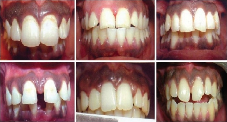
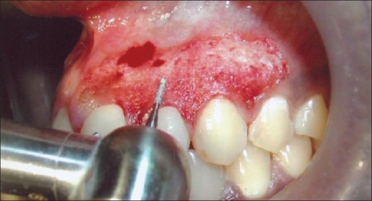
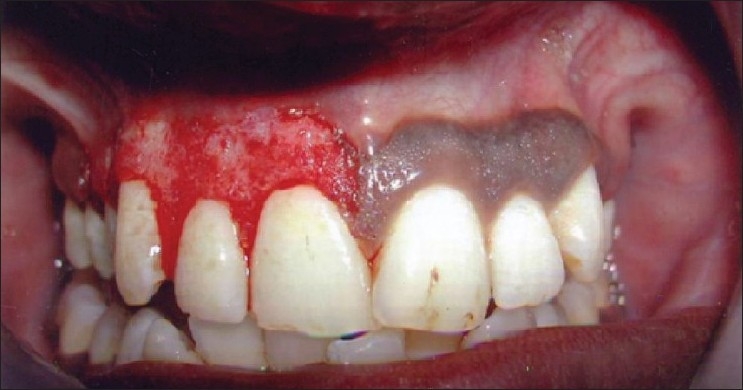
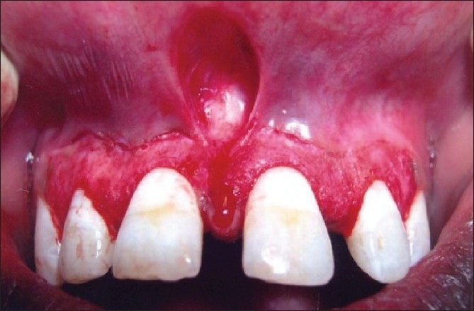
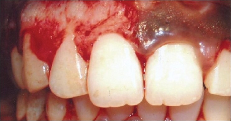
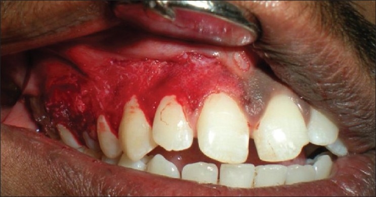
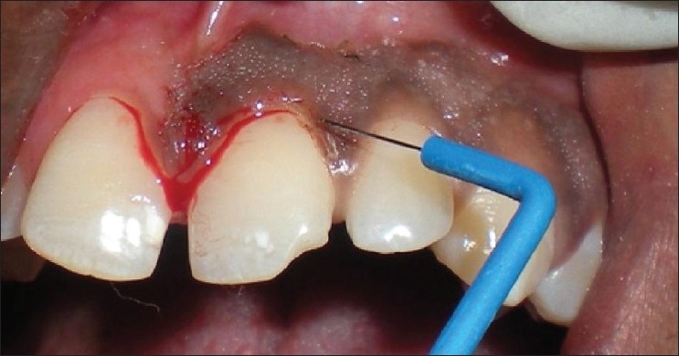
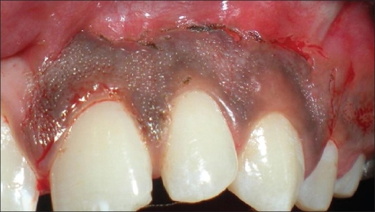
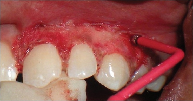
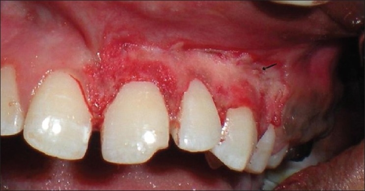
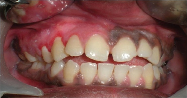
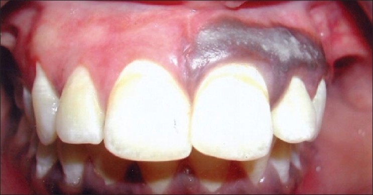
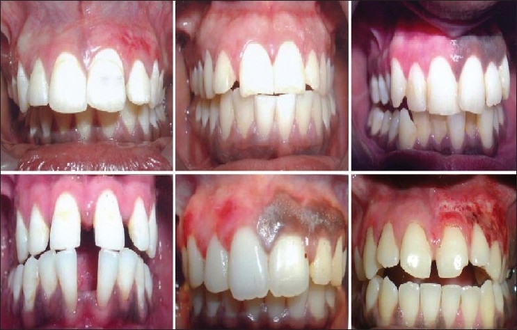
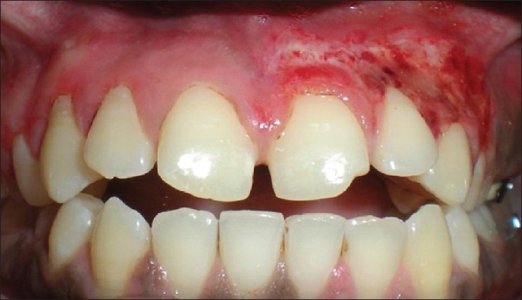
Gingival melanin pigmentation occurs in all races of mankind. Although clinical melanin pigmentation does neither present itself as a medical problem nor a disease entity, it is a major esthetic concern for many people, especially Asians. Esthetic gingival depigmentation procedures can be performed in such patients with excellent results. This case series presents a split mouth de-epithelization procedure using popular surgical techniques such as scalpel, bur abrasion or electrosurgery. These techniques were successfully used to treat gingival hyperpigmentation. Although we found that electrosurgery increased the efficacy of our work, giving a cleaner and neater work field, it required a lot of precision. In contrast, scalpel de-epithelization was easy and technique-friendly, giving excellent results and patient satisfaction. However, the cases are being followed-up to study the factors affecting the rate and length of time required for repigmentation and to study the repigmentation patterns. This case series also reviews the advantages and disadvantages of various techniques available for depigmentation, and reiterates that the scalpel technique still serves as a gold standard for depigmentation.
Keywords: Bur abrasion; depigmentation; electrosurgery; gingiva; melanin; physiological pigmentation; scalpel technique.
Conflict of interest statement
Figures
References
-
- Tal H, Oegiesser D, Tal M. Gingival depigmentation by Erbium: YAG laser: Clinical observations and patients responses. J Peroiodontol. 2003;74:1660–7. - PubMed
-
- Dummett CO. Oral pigmentation: First symposium of oral pigmentation. J Periodontol. 1960;31:356.
-
- Dummett CO, Barens G. Pigmentation of the oral tissues: A review of literature. J Periodontol. 1967;38:369–78. - PubMed
-
- Page LR, Corio RL, Crawford BE, Giansanti JS, Weathers DR. The Oral melanotic macule. Oral Surg Oral Med Oral Pathol. 1977;44:219–26. - PubMed
-
- Cicek Y. The normal and pathological pigmentation of oral mucous membrane: A review. J Contemp Dent Pract. 2003;4:76–86. - PubMed
Publication types
LinkOut - more resources
Full Text Sources
Other Literature Sources