. 2011:17:2440-54.
Epub 2011 Sep 21.

The chicken cornea as a model of wound healing and neuronal re-innervation

Affiliations

The chicken cornea as a model of wound healing and neuronal re-innervation

Eric R Ritchey et al. Mol Vis. 2011.

Abstract

Purpose: The cornea is the major refractive component of the eye and serves as a barrier to the external environment. Understanding how the cornea responds to injury is important to developing therapies to treat vision disorders that affect the integrity and refractive properties of the cornea. Thus, investigation of the wound healing responses of the cornea to injury in a cost-effective animal model is a valuable tool for research. This study characterizes the wound healing responses in the corneas of White Leghorn chicken.

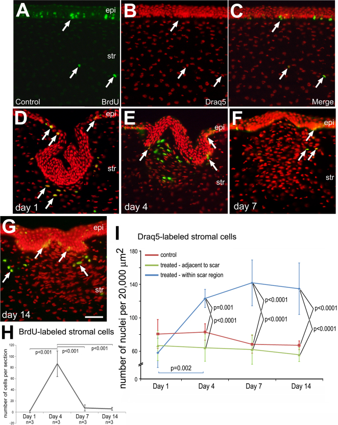
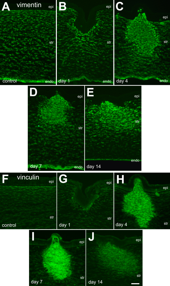
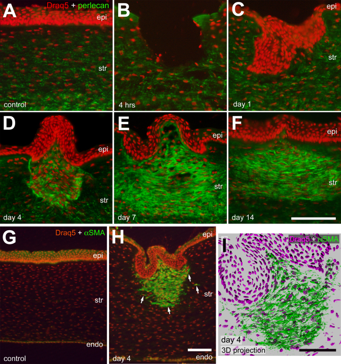
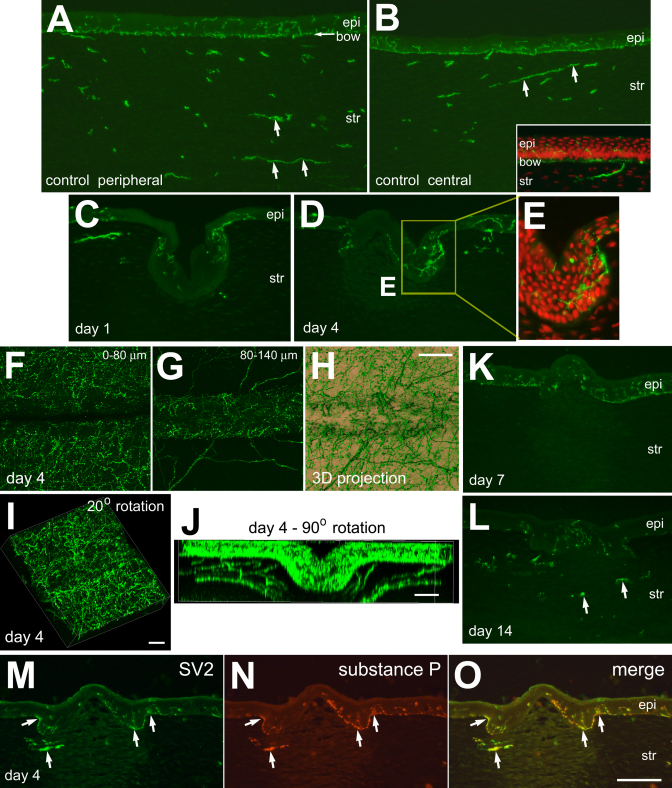
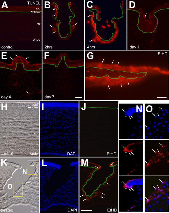
Methods: Linear corneal wounds were induced in post-natal day 7 (P7) chicks and cellular proliferation, apoptosis and regulation of structural proteins were assessed using immunohistochemical techniques. We describe the time course of increased expression of different scar-related markers, including vimentin, vinculin, perlecan and smooth muscle actin.

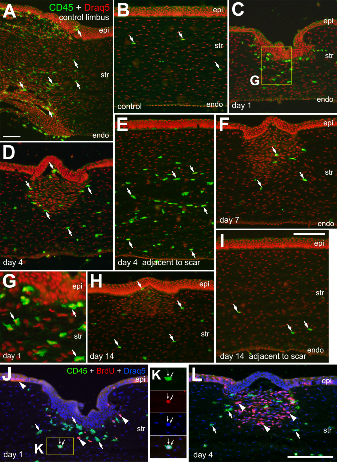
Results: We find evidence for acute necrotic cell death in the corneal region immediately surrounding cite of incision, whereas we failed to find evidence of delayed cell death or apoptosis. We find that the neuronal re-innervation of SV2-positive axon terminals within the corneal stroma and epithelium occurs very quickly after the initial scarring insult. We describe an accumulation of cells within the stroma immediately underlying the scar, which results, at least in part, from the local proliferation of keratocytes. Further, we provide evidence for scar-induced accumulations of CD45-positive monocytes in injured corneas.

Conclusions: We conclude that the chick cornea is an excellent model system in which to study wound healing, formation of scar tissue, and neuronal re-innervation of sensory endings.


Figures

Figure 1
Figure 1
Corneal injury stimulates the proliferation of stromal cells. Transverse sections of the cornea were labeled for BrdU (green) and Draq5 (red nuclei). Uninjured corneas (A-C) and injured corneas were harvested (and BrdU injected) at day 1 (BrdU injected 4 h prior; D), day 4 (BrdU injected at days 1, 2, and 3; E), day 7 (BrdU injected at days 4 and 5; F), and day 14 (BrdU injected at days 7 and 8; G). Arrows indicate BrdU-labeled nuclei. H: Plot indicating the mean (±SD; n=3 for each time point) number of BrdU-labeled cells within 20,000 µm2 within the stroma within the region of the incision. The calibration bar (50 µm) in G applies to A-G. I: Plots indicating the mean (±SD; n≥5 for each time point) indicate the mean number of stromal nuclei per 20,000 µm2 in control, undamaged corneas (red), treated stromal region adjacent to the incision site (green), and treated stromal region within the incision site (blue). ANOVA was performed to determine significance of difference within the data set and a Bonferroni post-hoc comparison was performed to determine significance between and within experimental groups. There was no significant difference (p>0.05) over time among treatment groups, with the exception of between 1 and 4 days after injury within the site of incision (p=0.002; I). Abbreviations: epi – epithelium, str – stroma, endo – endothelium.
Figure 2
Figure 2
The filamentous proteins vimentin and vinculin are upregulated in stromal cells following corneal injury. Transverse sections through the cornea were labeled with antibodies to vimentin (A-E) or vinculin (F-J). Corneas were harvested from control eyes (A and F) or 1 day (B and G), 4 days (C and H), 7 days (D and I) and 14 days (E and J) after injury. The calibration bar (50 µm) in J applies to all panels. Abbreviations: epi – epithelium, str – stroma, endo – endothelium.
Figure 3
Figure 3
α-Smooth muscle actin (αSMA) and perlecan are upregulated in the stroma following corneal injury. Transverse sections of the control (A and G) and treated (B-F, H and I) corneas were labeled with Draq5 (red nuclei) and antibodies perlecan (A-F; green) or αSMA (G-I; green). Corneas were harvested from control eyes (A and G) or 4 h (B), 1 day (C), 4 days (D, H, and I), 7 days (E), and 14 days (F) after injury. I is a 3D shadow reconstruction of a z-series of confocal optical sections to demonstrate the horizontal arrangement of presumptive myofibroblasts in the stroma within injured region. Arrows indicate cells labeled for αSMA and Draq5. The calibration bar (50 µm) in F applies to A-F, the bar in H applies to G and H, and the bar in I applies to I alone. Abbreviations: epi – epithelium, str – stroma, endo – endothelium.
Figure 4
Figure 4
CD45+ monocytes accumulate in the stroma following corneal injury. Transverse section of undamaged (A and B) and injured (C-L) corneas were labeled with Draq5 (red nuclei in A-I; blue nuclei J and L) and antibodies to CD45 (green) and BrdU (red in J and L). Corneas were harvest at 1 (C, G, and J), 4 (D, E, and L), 7 (F), and 14 (H and I) days after injury. Panel G is a 2.5 fold enlargement of the area (yellow box) in C indicating the amoeboid morphology of CD45+ monocytes in the site of injury in the stroma. Panel K is a 1.5 fold enlargement of the area (yellow box) in J providing a single-channel images and a merged overlay of labeling for CD45, BrdU and Draq5 in a single monocyte. Arrows indicate cells labeled for CD45 and Draq5, arrowheads indicate BrdU-labeled cells (J and L), and small double-arrows indicate a single cell labeled for CD45, BrdU and Draq5 (J and K). The calibration bar (50 µm) in panel A applies to A alone, the bar in I applies to B-I, and the bar in L applies to J and L. Abbreviations: epi – epithelium, str – stroma, endo – endothelium.
Figure 5
Figure 5
Neuronal re-innervation of the corneal epithelium occurs rapidly after injury. Transverse sections (A-E, H-O) or whole-mount preparations (F-J) were labeled with Draq5 (red) or antibodies to SV2 (green) and substance P (red; N and O). The inset in B is a high magnification view of SV2-positive neuronal ending in the basal layers of the corneal epithelium. Panel E is a twofold enlargement of the boxed-out (yellow rectangle) area in D. Panel F is a projection of confocal Z-stack of images taken from the surface of the epithelium down to 80 µm of depth. Panel G is a projection of confocal Z-stack of images from 80 µm down to 140 µm of depth from the surface of the cornea. Panel H is a 3D shadow reconstruction to demonstrate the arrangement of SV2-positive nerve terminals re-innervating regenerated epithelium. Panels I and J are 20° and 90° rotations of confocal Z-stack reconstructions to demonstrate the re-innervation of the trough of regenerated epithelium within the corneal incision. Arrows indicate SV2/substance P-positive axons in the stroma. The calibration bar (50 µm) in panel O applies to panels A-D, K, L, and M-O. The bar in H applies to all F-H panels, the bar in I applies to I alone, the bar in J applies to J alone. Abbreviations: epi – epithelium, str – stroma, endo – endothelium.
Figure 6
Figure 6
Corneal incision induces the death of epithelial and stromal cells shortly after injury. Transverse sections of the cornea were labeled for TUNEL (A-F), DAPI (I, L, N, and O), or EtHD (J, M, N, and O). Corneas were obtained at 15 min (G and K-O), 2 h (B), 4 h (C), 1 day (D), 4 days (E) or 7 days (F) after injury. The green lines indicate Bowman’s membrane and the edge of the incision. Panel G is a whole-mount preparation of an acutely injured cornea that was labeled with EtHD. Panels H and K are DIC images of control and damaged corneas. Panels N and O are 2.5 fold enlargements of the boxed-out (yellow rectangles) areas in panel K. Arrows indicate the nuclei of dying cells. The calibration bar (50 µm) in panel F applies to panels A-F, the bar in panel in G applies to G alone and the bar in panel M applies to panels H-M. Abbreviations: epi – epithelium, str – stroma, endo – endothelium.

References

    1. Hay ED. Development of the vertebrate cornea. Int Rev Cytol. 1980;63:263–322. - PubMed
    1. Zieske JD. Corneal development associated with eyelid opening. Int J Dev Biol. 2004;48:903–11. - PubMed
    1. Fini ME. Keratocyte and fibroblast phenotypes in the repairing cornea. Prog Retin Eye Res. 1999;18:529–51. - PubMed
    1. Wilson SE, Mohan RR, Ambrosio R, Jr, Hong J, Lee J. The corneal wound healing response: cytokine-mediated interaction of the epithelium, stroma, and inflammatory cells. Prog Retin Eye Res. 2001;20:625–37. - PubMed
    1. Jester JV, Petroll WM, Barry PA, Cavanagh HD. Expression of alpha-smooth muscle (alpha-SM) actin during corneal stromal wound healing. Invest Ophthalmol Vis Sci. 1995;36:809–19. - PubMed

Publication types

MeSH terms