. 2011 Sep;7(9):e1002267.
doi: 10.1371/journal.ppat.1002267. Epub 2011 Sep 22.

Exposure to the viral by-product dsRNA or Coxsackievirus B5 triggers pancreatic beta cell apoptosis via a Bim / Mcl-1 imbalance

Affiliations

Exposure to the viral by-product dsRNA or Coxsackievirus B5 triggers pancreatic beta cell apoptosis via a Bim / Mcl-1 imbalance

Maikel L Colli et al. PLoS Pathog. 2011 Sep.

Abstract

The rise in type 1 diabetes (T1D) incidence in recent decades is probably related to modifications in environmental factors. Viruses are among the putative environmental triggers of T1D. The mechanisms regulating beta cell responses to viruses, however, remain to be defined. We have presently clarified the signaling pathways leading to beta cell apoptosis following exposure to the viral mimetic double-stranded RNA (dsRNA) and a diabetogenic enterovirus (Coxsackievirus B5). Internal dsRNA induces cell death via the intrinsic mitochondrial pathway. In this process, activation of the dsRNA-dependent protein kinase (PKR) promotes eIF2α phosphorylation and protein synthesis inhibition, leading to downregulation of the antiapoptotic Bcl-2 protein myeloid cell leukemia sequence 1 (Mcl-1). Mcl-1 decrease results in the release of the BH3-only protein Bim, which activates the mitochondrial pathway of apoptosis. Indeed, Bim knockdown prevented both dsRNA- and Coxsackievirus B5-induced beta cell death, and counteracted the proapoptotic effects of Mcl-1 silencing. These observations indicate that the balance between Mcl-1 and Bim is a key factor regulating beta cell survival during diabetogenic viral infections.


Conflict of interest statement

The authors have declared that no competing interests exist.

Figures
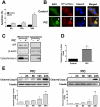
Figure 1
Figure 1. Internal dsRNA induces beta cell death via the mitochondrial pathway of apoptosis.
A. INS-1E cells were transfected with the synthetic dsRNA polyinosinic-polycytidylic acid (PIC) and apoptosis was evaluated at different time points using nuclear dyes as described in Methods (n = 5, *P<0.01 vs. untreated). B. BAX localization was studied by immunocytochemistry in INS-1E cells exposed or not to internal dsRNA for 24h. Pictures are representative of 4 independent experiments; arrows indicate BAX mitochondrial localization, similar to the ATP Synthase (mitochondrial marker) C. Cytochrome c release in INS-1E cells treated for 24h in the presence or absence of intracellular dsRNA. Pictures are representative of 4 independent experiments. D. Western blot quantified by densitometry of cytoplasmic protein fractions in INS-1E cells transfected or not with dsRNA for 24h (n = 4, *P<0.01 vs. untreated). E . Western blot analysis and densitometry of cleaved caspases 9 and 3 after INS-1E cells exposure to internal dsRNA at different time points (n = 5–6, *P<0.05, **P<0.01 vs. untreated). Data are mean ± SEM.
Figure 2
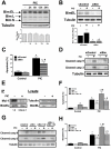
Figure 2. Mcl-1 protein expression is decreased by internal dsRNA and its modulation regulates beta cell apoptosis.
A. INS-1E cells were transfected with dsRNA for different time points and Mcl-1 expression was analyzed by Western blot (top) and quantified by densitometry (bottom; data are shown as fold-change compared to non-treated cells; -) (n = 3, *P<0.01 vs. untreated). B. mRNA expression of Mcl-1 in INS-1E cells treated or not with internal dsRNA for 24h (n = 5, *P<0.01 vs. untreated). Results are corrected for the housekeeping gene GAPDH. C. INS-1E cells were treated or not for 12h with the proteasome inhibitor MG-132 in the presence or absence of intracellular PIC and Mcl-1 expression was analyzed by Western blot (top) and quantified by densitometry (bottom; data are shown as fold-change compared to non-treated cells; -) (n = 4, *P<0.01 vs. control (untreated), #P<0.05 vs. PIC alone). D, E and F. INS-1E cells (D and E) or FACS-purified primary beta cells (F) were transfected with siControl or with two different small-interfering RNAs against Mcl-1 (siMcl-1 and siMcl-1 #2) for 48h, as described in Methods. D. Protein expression of Mcl-1 and cleaved caspases 9 and 3 was evaluated by Western blot. Pictures are representative of 3 independent experiments. E and F. dsRNA-induced apoptosis in INS-1E cells (E) and FACS-purified primary beta cells (F) treated for 24 h with PIC with or without Mcl-1 knockdown (n = 5, *P<0.01 vs. siControl, #P<0.01 vs. siControl + PIC). G, H and I. INS-1E cells (G and H) or FACS-purified primary beta cells (I) were infected or not (NI) with adenovirus encoding the luciferase gene (adLuc) or rat Mcl-1 (adMcl-1) at different multiplicities of infection (MOI) for 48h, as indicated. G. Protein expression of Mcl-1 or cleaved caspases 9 and 3 were evaluated by Western blot 24h after PIC exposure. Pictures are representative of 3 independent experiments. H and I. dsRNA-induced apoptosis in INS-1E cells (H) and FACS-purified primary beta cells (I) treated for 24h with PIC with or without Mcl-1 overexpression (n = 4, *P<0.01 vs. NI, #P<0.01 vs. NI + PIC). Data are mean ± SEM.
Figure 3
Figure 3. Bim contributes for dsRNA-induced apoptosis and its silencing prevents the proapoptotic effects of Mcl-1 knockdown.
A. Time course analysis of Bim protein expression in INS-1E cells treated with internal dsRNA. Cells were harvested at different time points and Bim expression was evaluated by Western blot (top) and quantified by densitometry (bottom) (n = 10). B to D. INS-1E cells were transfected with siControl or a specific siRNA against Bim (siBim) for 24 h and then exposed to intracellular dsRNA for 24h. Bim knockdown was confirmed by Western blot (B). Cell viability (C) was measured using nuclear dyes (n = 3, *P<0.01 vs. siControl, #P<0.05 vs. siControl + PIC). Activation of caspases 9 and 3 (D) was determined in the presence or not of Bim silencing. E. INS-1E cells were left untreated or transfected with dsRNA for 24h and then subjected to immunoprecipitation with an antibody against Bim (IP). The presence of Mcl-1 and Bim in the immunoprecipitated material was determined using specific antibodies, and the ratio Mcl-1/Bim band intensity determined by densitometry. The total lysate (Lysate) was evaluated by Western blotting with anti-Bim and anti-α-tubulin antibodies. The pictures shown are representative of 3–4 independent experiments. INS-1E cells (F and G) or FACS-purified primary beta cells (H) were transfected with siControl, siBim, siMcl-1 or a combination of siBim plus siMcl-1. After a 24h recovery period they were then exposed to internal dsRNA for 24 h. Cell viability (F and H) and cleavage of caspases 9 and 3 (G) were determined after 24h exposure to dsRNA (n = 3 - 5, *P<0.01 vs. siControl, #P<0.05 vs. siControl + PIC, ##P<0.01 vs. siControl + PIC, §P<0.01 vs. siMcl-1 + PIC). The pictures shown are representative of 3 independent experiments. Data are mean ± SEM.
Figure 4
Figure 4. CVB5 infection induces apoptosis in beta cells via the BH3-only protein Bim.
A to G. INS-1E cells (A to E) or FACS-purified primary beta cells (F and G) were infected with CVB5 at the indicated MOIs, as described in Methods. Protein expression of the viral capsids was evaluated by immunocytochemistry at 24h using an enterovirus-specific antibody (A). Cell apoptosis (B) and caspases 9 and 3 cleavage (C) were determined in cells infected or not with CVB5 for 24 h. (n = 3, *P<0.01 vs. uninfected, §P<0.01 vs. MOI 5). D to G. INS-1E cells (D) or primary beta cells (G) were transfected with siControl or a specific siRNA against Bim (siBim) for 24 h and then infected with CVB5 at the MOI 5. Cell viability was determined after 18h (D and G) (n = 3, *P<0.01 vs. siControl, #P<0.05 vs. siControl + PIC). Bim knockdown, caspases 9 and 3 cleavage and the expression of the viral capsids (VP1 and 2) were evaluated by Western blot (E and F). The pictures shown are representative of 3 independent experiments. Data are mean ± SEM.
Figure 5
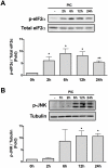
Figure 5. Internal dsRNA induces eIF2α and JNK phosphorylation.
Cell lysates of INS-1E cells transfected with dsRNA for different time points were used for Western blotting with antibodies against p-eIF2α (A) or p-JNK (B). Densitometry was performed and the results were normalized for the expression of total eIF2α (A) or α-tubulin (B) (n = 5, *P<0.01 vs. untreated, **P<0.05 vs. untreated). The pictures shown are representative of 5 independent experiments. Data are mean ± SEM.
Figure 6
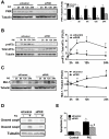
Figure 6. dsRNA-induced decrease in Mcl-1 protein expression depends on eIF2α phosphorylation by PKR.
A to E. Cells were transfected with siControl or a siRNA against PKR (siPKR) and 48h after recovery treated with internal dsRNA for different time points. The expression of PKR (A), p-eIF2α (B) and Mcl-1 (C) was determined by Western blot (left) and quantified by densitometry (right) in INS-1 E cells (n = 4, *P<0.01 vs. siControl + PIC at the same time point). Cleavage of caspase 9 and 3 (D) and cell viability (E) were evaluated after 24h of dsRNA exposure with or without siPKR (n = 5, *P<0.01 vs. siControl, **P<0.05 vs. siControl, #P<0.01 vs. siControl + PIC). The pictures shown are representative of 4-5 independent experiments. Data are mean ± SEM.
Figure 7
Figure 7. Schematic representation of dsRNA- and virus-induced beta cell apoptosis.
During viral infection of beta cells the viral by-product dsRNA is released into the cytoplasm. 1) Cytoplasmic dsRNA activates the kinase PKR that in turn phosphorylates the elongation factor eIF2α; 2) phosphorylation of eIF2α leads to inhibition of protein translation with consequent decrease of Mcl-1 expression; 3) reduction of Mcl-1 protein increases unbound Bim, which activates BAX; 4) BAX translocates to the mitochondria; 5) BAX oligomerizes and induces permeabilization of the mitochondrial outer membrane, allowing the release of cytochrome c to the cytoplasm; 6) cytoplasmic cytochrome c forms complexes with Apaf1 and activates the initiator caspase 9; 7) finally, caspase 9 cleaves and activates the executioner caspase 3, 8) triggering the final steps of beta cell apoptosis; 9) further studies are required to clarify additional pathways implicated in virus-induced beta cell apoptosis.

Similar articles

Cited by

References

    1. Eizirik DL, Colli ML, Ortis F. The role of inflammation in insulitis and beta cell loss in type 1 diabetes. Nat Rev Endocrinol. 2009;5:219–226. - PubMed
    1. Tao B, Pietropaolo M, Atkinson M, Schatz D, Taylor D. Estimating the cost of type 1 diabetes in the U.S.: a propensity score matching method. PLoS One. 2010;5:e11501. - PMC - PubMed
    1. Concannon P, Rich SS, Nepom GT. Genetics of type 1A diabetes. N Engl J Med. 2009;360:1646–1654. - PubMed
    1. Colli ML, Moore F, Gurzov EN, Ortis F, Eizirik DL. MDA5 and PTPN2, two candidate genes for type 1 diabetes, modify pancreatic beta cell responses to the viral by-product double-stranded RNA. Hum Mol Genet. 2010;19:135–146. - PMC - PubMed
    1. Patterson CC, Dahlquist GG, Gyurus E, Green A, Soltesz G. Incidence trends for childhood type 1 diabetes in Europe during 1989-2003 and predicted new cases 2005-20: a multicentre prospective registration study. Lancet. 2009;373:2027–2033. - PubMed

Publication types

MeSH terms