Pulmonary alveolar microlithiasis with low fluorodeoxyglucose accumulation in PET/computed tomography
- PMID: 21977072
- PMCID: PMC3183644
- DOI: 10.4103/1817-1737.84781
Pulmonary alveolar microlithiasis with low fluorodeoxyglucose accumulation in PET/computed tomography
Abstract
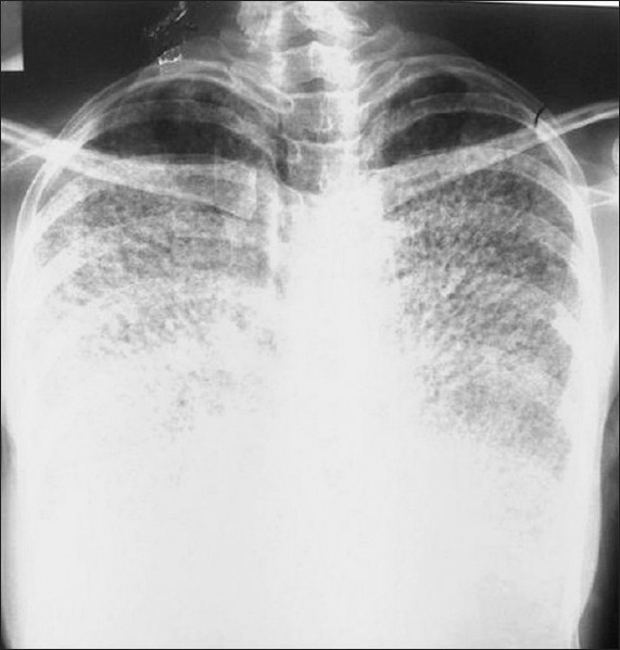
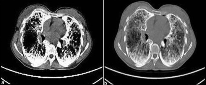
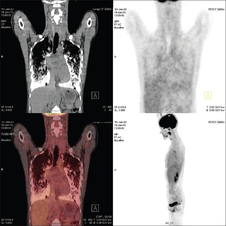
Pulmonary alveolar microlithiasis (PAM) is an uncommon lung disease characterized by accumulation of intraalveolar calcifications. The disease can be diagnosed based on the radiological findings. We present a 27-year-old women with five-year history of shortness of breath. She was diagnosed with PAM due to the presence of the characteristic chest X-ray and thorax computed tomography (CT) findings. We performed (18)F-fluorodeoxyglucose (FDG)-PET/CT imaging in order to detect any evidence of inflamation in the lung before deciding an anti-inflammatory treatment. The lung regions with dense calcifications revealed low FDG uptakes (SUVmax: 2.7) and the lung regions without calcifications showed lower FDG uptakes. No further treatment modality was planned besides inhaler salbutamol. Herein, we discuss this rare entity with literature search.
Keywords: FDG; PET/CT; inflammation; pulmonary alveolar microlithiasis.
Conflict of interest statement
Figures
References
-
- Friedrich N. Corpora amylacea in den lungen. Arch Pathol Anat. 1856;9:613–8.
-
- Harbitz F. Extensive calcifications of the lungs as distinct disease. Arch Intern Med. 1918;21:139–46.
-
- Ito K, Kubota K, Yukihiro M, Izumi S, Miyano S, Kudo K, et al. FDG-PET/CT finding of high uptake in pulmonary alveolar microlithiasis. Ann Nucl Med. 2007;21:415–8. - PubMed
-
- Castellana G, Lamorgese V. Pulmonary alveolar microlithiasis world causes and review of the literature. Respiration. 2003;70:549–55. - PubMed
-
- Deniz O, Ors F, Tozkoparan E, Ozcan A, Gumus S, Bozlar U, et al. High resolution computed tomographic features of pulmonary alveolar microlithiasis. Eur J Radiol. 2005;55:452–60. - PubMed
Publication types
LinkOut - more resources
Full Text Sources
Miscellaneous